All of the evidence provided on Hippocampome.org are quotes from scientific texts.
However, because quoted passages may be difficult to understand in isolation, contextual information and expanded abbreviations set in square brackets have been added for clarity.
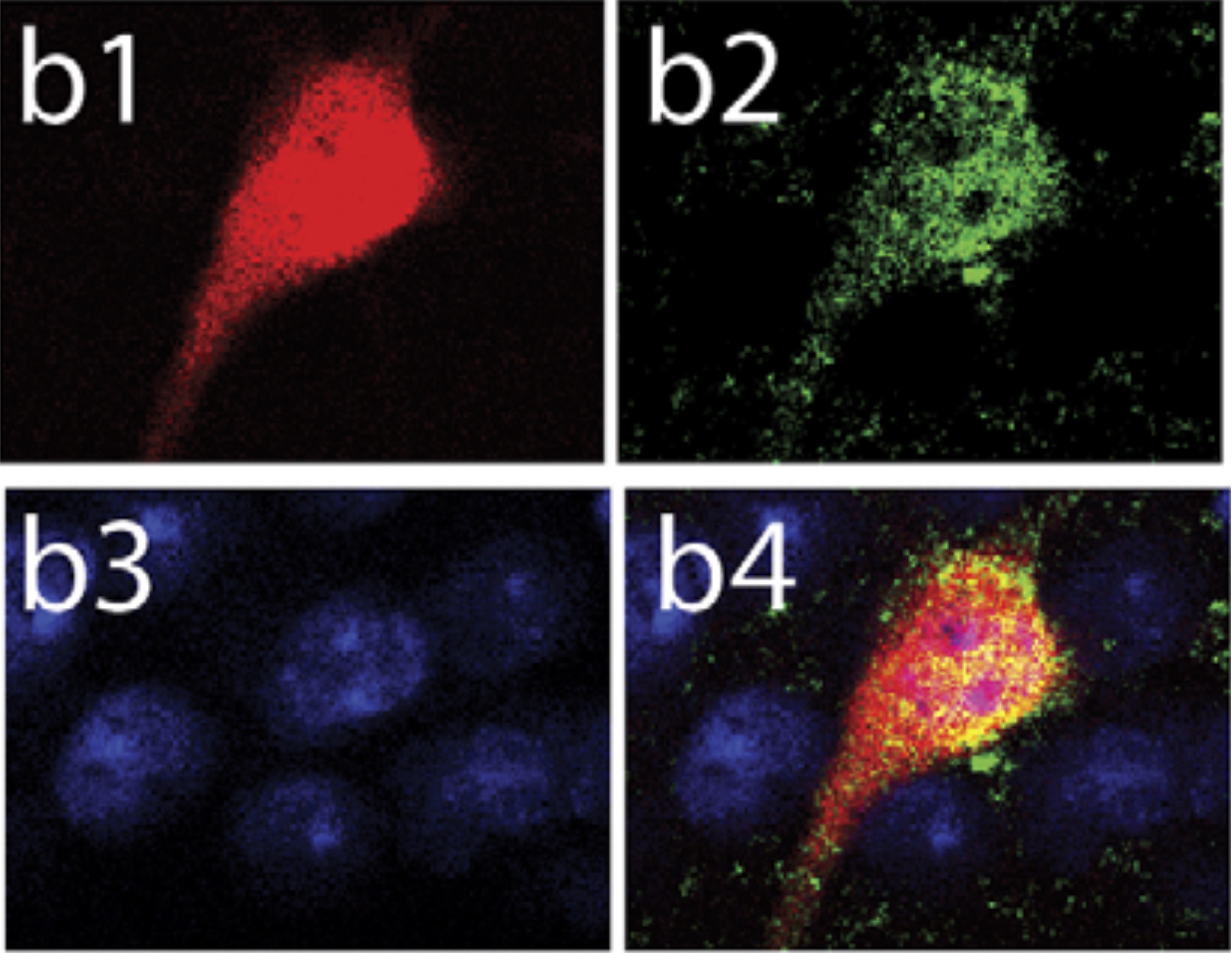
"FIGURE 1. Characterization of hippocampal CRH [corticotropin-releasing hormone] neurons. a, A representative image of the hippocampus from Crh-GFP [green fluorescent protein] mice. a1, Higher magnification of the CA1 pyramidal cell layer highlights the morphology of hippocampal CRH neurons. a2, Higher magnification of the CA3 subregion of the hippocampus reveals dense GFP-expressing terminals surrounding the principal neurons. b, Representative image of a tdTomato-expressing hippocampal CRH neurons (red; b1) that is also immunoreactive for CRH (green; b2) (n = 232 neurons, four mice)."
 Browse
 Search
 Help
 positive