All of the evidence provided on Hippocampome.org are quotes from scientific texts.
However, because quoted passages may be difficult to understand in isolation, contextual information and expanded abbreviations set in square brackets have been added for clarity.
Animal:
Protocol:
Expression:
Show:
EXPRESSION: positive
ANIMAL: mouse
PROTOCOL: promoter_expression_construct
Page location: p2863, Fig. 1
Linking PMID: 20547684 Linking Quote: All necessary morphological evidence is present in this self-same publication. Neurons in CA1 with the Bistratified axo-dendritic pattern of both axons and dendrites in only SR, SP, and SO that are parvalbumin negative are considered to be CA1 0333 Ivy (see Fuentealba 2008 PMID-18367092, p919 left bottom). "Although all neurons in this study were fast spiking, only basket cells were immunoreactive for parvalbumin, suggesting that parvalbumin is not related to the fast neuronal discharge. [p6663, right top]"
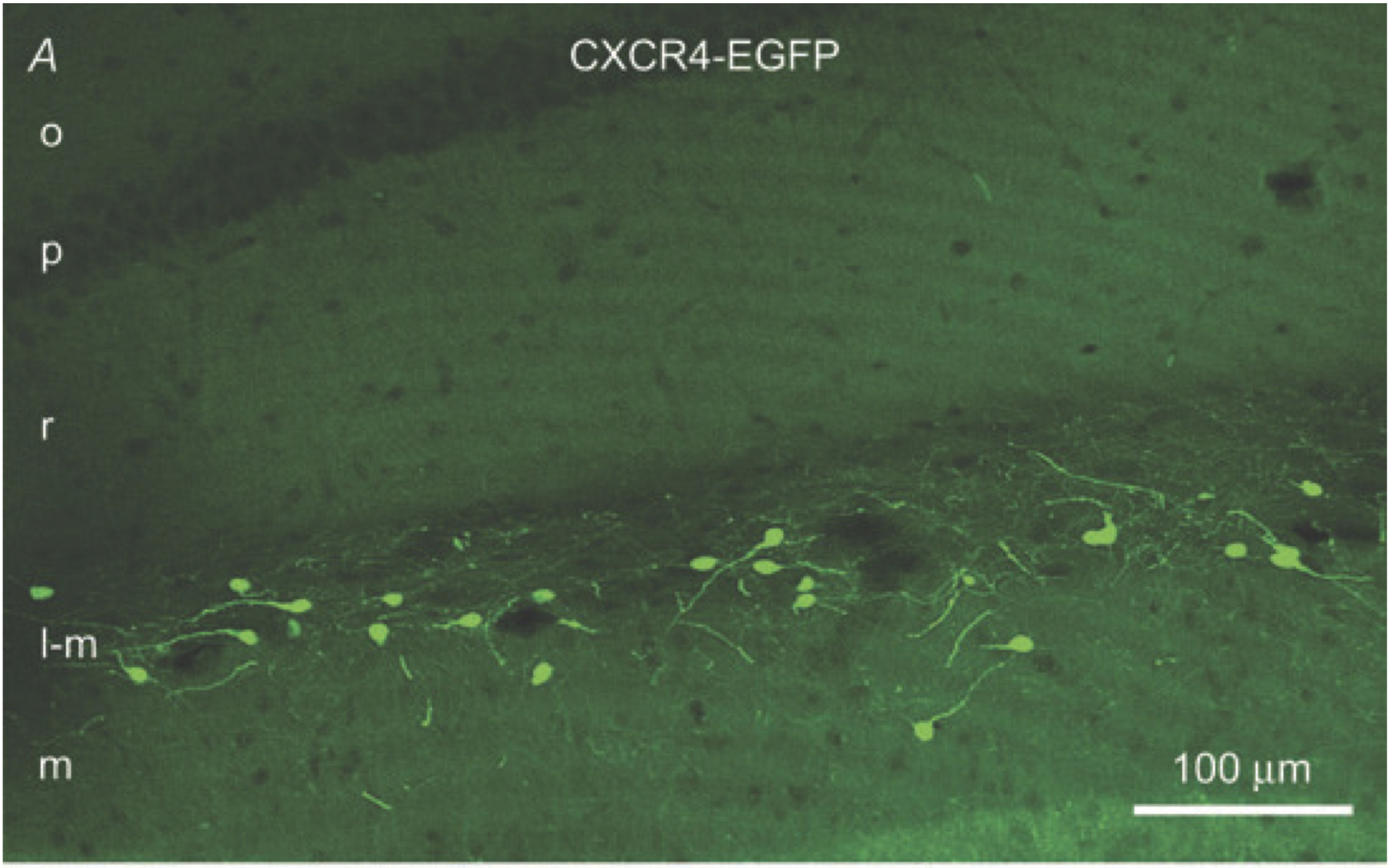
"Figure 1. Confocal ?uorescence microscopy demonstrates EGFP-identi?ed Cajal_Retzius cells in the CA1
hippocampus of a P24 CXCR4-EGFP mouse
A, EGFP-labelled cells situated along the hippocampal ?ssure in the stratum lacunosum-moleculare of CA1 and
the molecular layer of the fascia dentata have morphological features of previously demonstrated Cajal_Retzius
cells. Most of these neurons have a single dendrite that extends opposite to the axon initial segment
and that is oriented in different directions. The axons of the EGFP-labelled cells form a dense network in
the stratum lacunosum-moleculare. . . . B, this row of panels shows that all EGFP-labelled cells colocalize
reelin, which is enriched at the base of the single dendrite (arrowhead). Reelin-positive/EGFP-negative cells are
putative interneurons (
?
). C, this row of panels indicates that EGFP-positive cells also express calretinin, which
highlights the density of the axonal plexus of Cajal_Retzius cells."
 Browse
 Search
 Help
 positive