All of the evidence provided on Hippocampome.org are quotes from scientific texts.
However, because quoted passages may be difficult to understand in isolation, contextual information and expanded abbreviations set in square brackets have been added for clarity.
Animal:
Protocol:
Expression:
Show:
Order:
EXPRESSION: negative (inference)
ANIMAL: rat
PROTOCOL: immunohistochemistry
Page location: p1806, right middle
"All PV[parvalbumin]+ neurons [in CA3 and CA1] were positive for the GABA[gamma aminobutyric acid]A receptor [alpha]1 subunit [reference 31] (Fig._5b-d)."
Interpretation notes: Glutamatergic and GABAergic GABAa alpha1- types in CA1 SLM_SR_SP_SO are probably PV- by contrapositive logic
EXPRESSION: negative (inference)
ANIMAL: mouse
PROTOCOL: immunohistochemistry
Page location: p49, Table 2
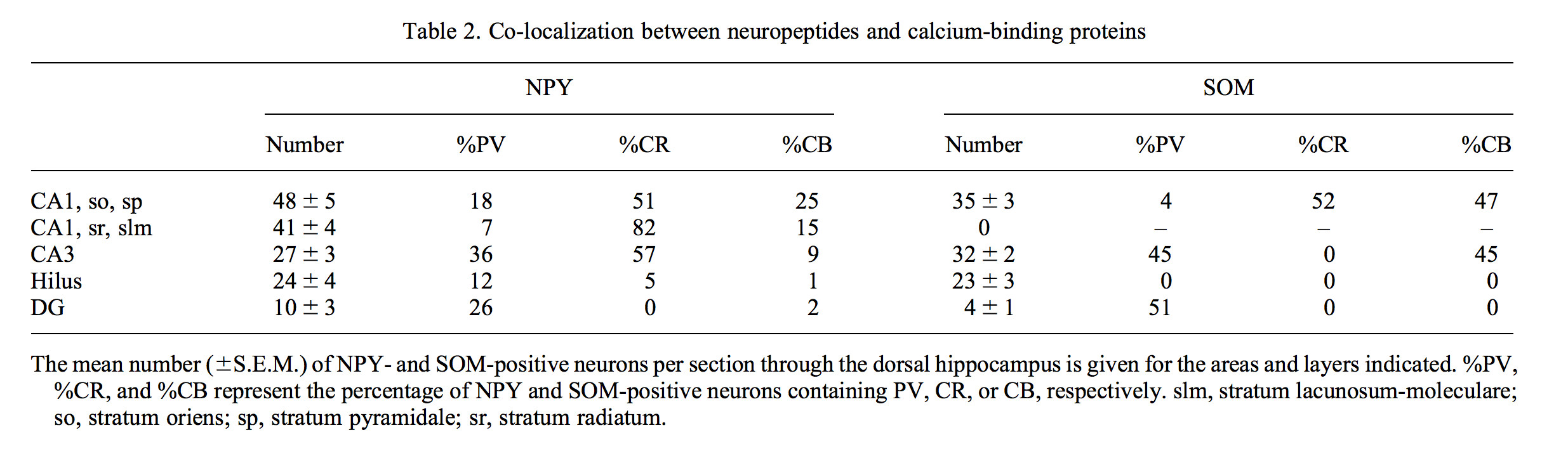
"Table 2. Co-localization between neuropeptides and calcium-binding proteins"
Interpretation notes: Glutamatergic and GABAergic NPY+ types in CA1 SP_SO are probably PV-; Clopper-Pearson confidence interval lower bound = 67.4% (n = 39/48)
EXPRESSION: negative (inference)
ANIMAL: mouse
PROTOCOL: immunohistochemistry
Page location: p49, Table 2
"Table 2. Co-localization between neuropeptides and calcium-binding proteins"
Interpretation notes: Glutamatergic and GABAergic SOM+ types in CA1 SP_SO are probably PV-; Clopper-Pearson confidence interval lower bound = 85.1% (n = 34/35)
EXPRESSION: negative (inference)
ANIMAL: rat
PROTOCOL: mRNA
Page location: p265, left mid
"In the hippocampus, cells expressing SOM [somatostatin], PARV [parvalbumin], and GAD67 [glutamic acid decarboxylase 67] were studied in the stratum oriens of the cornu ammonis 1 (CA1), the cornu ammonis 3 (CA3), and the dentate gyrus. All neurons labeled by the digoxygenin probes in both hemispheres of three different rats were studied. A total of 391 neurons were examined (Fig. 5). In CA1, labeling for mGluR1 was found in cells expressing SOM (P<0.0001) while no significant labeling was observed in the cells expressing GAD67 or PARV. In CA3 and the dentate hilus, labeling for mGluR1 was seen strongly in cells expressing SOM and lightly in GAD67-expressing cells (P<0.0001), for both comparisons, but not for PARV-expressing cells. Fig. 6 . Other than the SOM-expressing neurons, there were no other intensely mGluR1 labeled cells seen in the stratum oriens of CA1 and CA3. There were strongly labeled neurons in the dentate hilus that did not express SOM."
Interpretation notes: Glutamatergic and GABAergic mGluR1+ types in CA1 SO are probably PV-
EXPRESSION: negative (inference)
ANIMAL: rat
PROTOCOL: immunohistochemistry
Page location: p841, Table 2
"TABLE 2. Proportion of neurons stained for GABA, calcium-binding proteins
and neuropeptides expressing the a1 subunit-immunoreactivity "
Interpretation notes: Glutamatergic and GABAergic GABAa alpha1- types in CA1 SLM_SR_SP_SO are probably PV- by contrapositive logic
 Browse
 Search
 Help
 negative; confirmed by inference(s)