All of the evidence provided on Hippocampome.org are quotes from scientific texts.
However, because quoted passages may be difficult to understand in isolation, contextual information and expanded abbreviations set in square brackets have been added for clarity.
Animal:
Protocol:
Expression:
Show:
EXPRESSION: positive (inference)
ANIMAL: rat
PROTOCOL: immunohistochemistry
Page location: p190. Fig. 3
Linking PMID: 19699779 Linking Quote: Principal neurons identified by the presence of their somata in the principal cell layer, which is densely packed with and nearly homogeneous for the somata of the subregion's principal neurons (Czeh et al., 2013, Acta Biologica Hungarica).
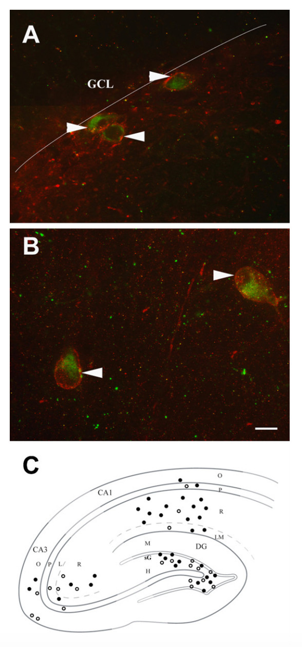
"Fig. 3. NK1 and VGLUT3 were colocalized in the hippocampal formation (A-C) but not in the median raphe nucleus (D). (A) A photomontage of three cells in the dentate gyrus that contain both NK1 outlining the cytoplasm (red) and VGLUT3 (green, arrow heads). The white line marks the junction between the granule cell layer (GCL) and the hilus, these cells are located just at the border in the subgranular layer. (B) Two neighboring interneurons located in stratum radiatum of the CA1 region are dually labeled for NK1 and VGLUT3 (arrowheads). Both panels same scale bar = 20 [micro]m. (C) Relative proportions of cells dually labeled for VGLUT3 and NK1 (filled circles) and cells containing only VGLUT3 (open circles) by laminar location and subfield, CA1, CA3 and dentate gyrus (DG). Several sections were sampled and single VGLUT3 labeled and cells dually labeled for VGLUT3 and NK1 were counted in each lamina. Medio-lateral distribution of cells is compressed and between subfield comparisons
are not quantitative since each region was sampled independently from a different number of sections. For the CA1 region, most cells containing VGLUT3 were located in stratum radiatum (86% or 66/77) and the majority of these also contained NK1 immunolabeling (89% or 59/66). In all the remaining areas, the percentage of VGLUT3 cells that also contained NK1 averaged 44-62%, except for stratum radiatum of the CA3 region where 71% were dually labeled. Total number of cells sampled in CA1 = 96, CA3 = 19, and DG = 120."
Interpretation notes: Glutamatergic and GABAergic vGluT3+ types in CA1 SR are probably Sub P rec+; Clopper-Pearson confidence interval lower bound = 79.4% (n = 59/66)
 Browse
 Search
 Help
 positive inference