All of the evidence provided on Hippocampome.org are quotes from scientific texts.
However, because quoted passages may be difficult to understand in isolation, contextual information and expanded abbreviations set in square brackets have been added for clarity.
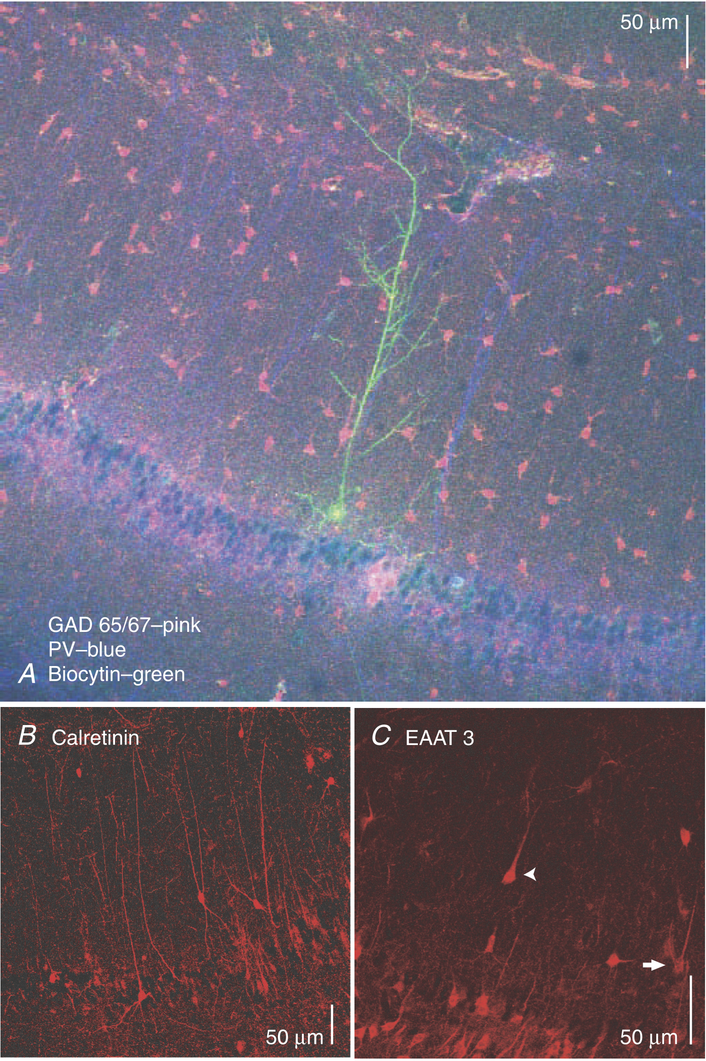
"Figure 2. [CA1] PLP [pyramidal-like principal] neurons do not stain for interneuronal markers A, PLPs were immunonegative for glutamic acid decarboxylase 65/67 (GAD 65/67; pink) and parvalbumin (PV; blue). GAD-positive interneurons were visible in all layers of CA1, in addition to the PV-positive dendrites of putative basket cells (blue). A biocytin-filled PLP neuron was stained with streptavidin/Alexa-488 (green). B, calretinin-positive interneurons were brightly stained, while neurons with PLP morphology were not visible. C, staining for excitatory amino acid transporter 3 (EAAT3) revealed an immunopositive PLP neuron (arrowhead) and a radiatum giant cell (RGC; arrow), along with numerous pyramidal neurons."
"We then incubated slices with an antibody for excitatory amino acid transporter 3 (EAAT3, EAAC1). Although known to be present on inhibitory neurons in the cerebellum, EAAT3 consistently stains pyramidal neurons in the hippocampus (Rothstein et al. 1994; Kanai et al. 1995). We found dense staining for both [CA1] pyramidal neurons and [CA1] neurons with PLP [pyramidal-like principal neuron] morphology in the [CA1] SR [stratum radiatum] (Fig. 2C, arrowhead). Also present were neurons with the typical 'radiatum giant cell' (RGC) morphology (Gulyas et al. 1998) as described below (Fig. 2C, arrow)."
"Figure 2. [CA1] PLP [pyramidal-like principal] neurons do not stain for interneuronal markers A, PLPs were immunonegative for glutamic acid decarboxylase 65/67 (GAD 65/67; pink) and parvalbumin (PV; blue). GAD-positive interneurons were visible in all layers of CA1, in addition to the PV-positive dendrites of putative basket cells (blue). A biocytin-filled PLP neuron was stained with streptavidin/Alexa-488 (green). B, calretinin-positive interneurons were brightly stained, while neurons with PLP morphology were not visible. C, staining for excitatory amino acid transporter 3 (EAAT3) revealed an immunopositive PLP neuron (arrowhead) and a radiatum giant cell (RGC; arrow), along with numerous pyramidal neurons."
 Browse
 Search
 Help
 positive