positive-negative: subcellular expression differences Explanation: Protein has been detected in dendrites, but not soma.
All of the evidence provided on Hippocampome.org are quotes from scientific texts.
However, because quoted passages may be difficult to understand in isolation, contextual information and expanded abbreviations set in square brackets have been added for clarity.
Animal:
Protocol:
Expression:
Show:
Order:
EXPRESSION: negative
ANIMAL: rat
PROTOCOL: immunohistochemistry
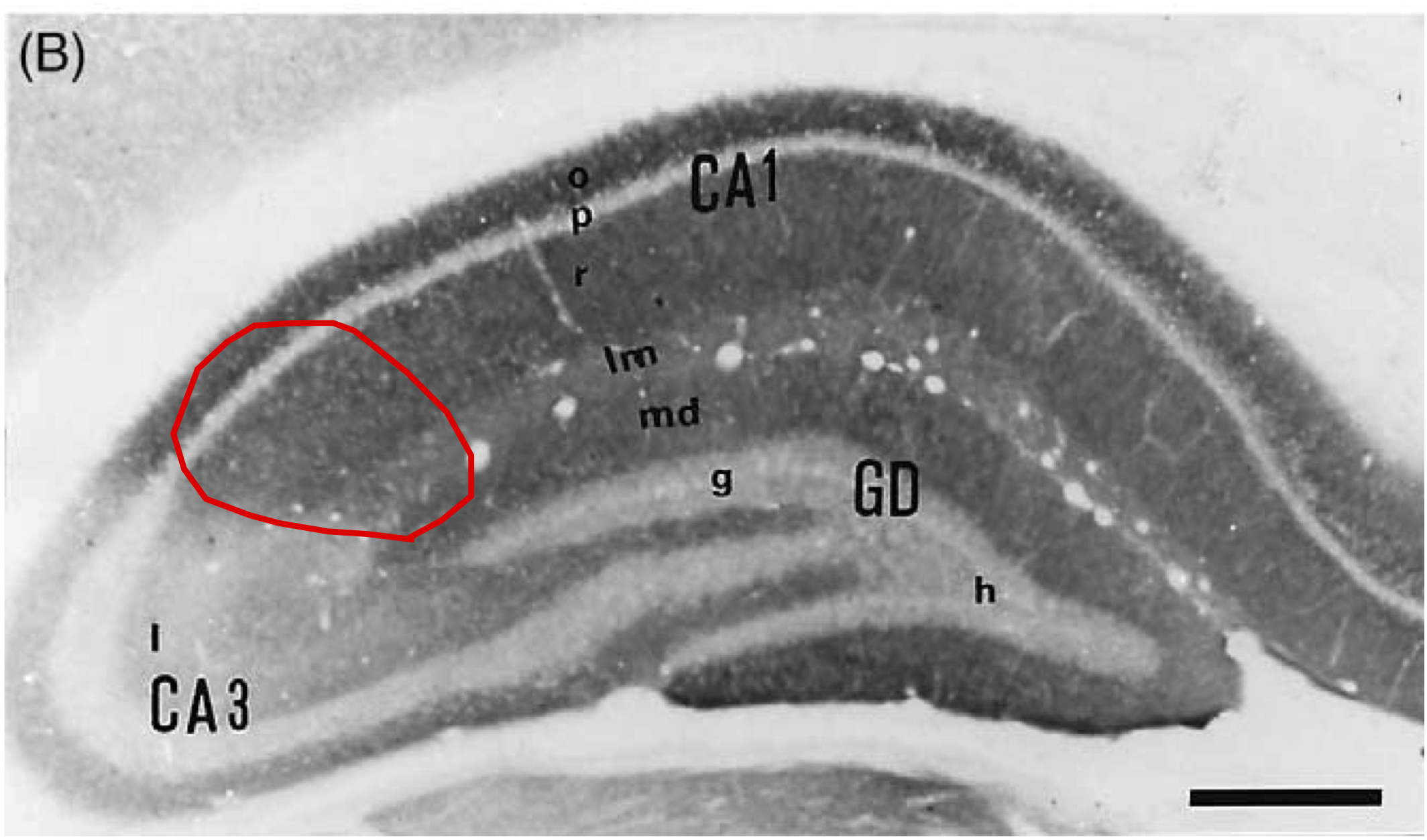
Page location: p1018, Fig. 1
Linking PMID: 12128002 Linking Quote: Principal neurons identified by the presence of their somata in the principal cell layer, which is densely packed with and nearly homogeneous for the somata of the subregion's principal neurons (Czeh et al., 2013, Acta Biologica Hungarica).
"Fig. 1. Distribution of immunoreactivity (IR) for mGluR1a (A) and
mGluR5a (B) in the rat dorsal hippocampus."
EXPRESSION: negative
ANIMAL: rat
PROTOCOL: immunohistochemistry
Page location: p1021, left top
Linking PMID: 12128002 Linking Quote: Principal neurons identified by the presence of their somata in the principal cell layer, which is densely packed with and nearly homogeneous for the somata of the subregion's principal neurons (Czeh et al., 2013, Acta Biologica Hungarica).
"The immunoreactivity of mGluR5 and also of its
splice variant mGluR5a was observed by us and other
authors in the neuropil, mainly on dendrites of principal
hippocampal neurons (Lujan et al., 1996; Shigemoto et
al., 1997)."
 Browse
 Search
 Help
 positive-negative: subcellular expression differences