All of the evidence provided on Hippocampome.org are quotes from scientific texts.
However, because quoted passages may be difficult to understand in isolation, contextual information and expanded abbreviations set in square brackets have been added for clarity.
Animal:
Protocol:
Expression:
Show:
EXPRESSION: negative
ANIMAL: mouse
PROTOCOL: immunohistochemistry
Page location: p124, Fig. 5
Linking PMID: 17942321 Linking Quote: Principal neurons identified by the presence of their somata in the principal cell layer, which is densely packed with and nearly homogeneous for the somata of the subregion's principal neurons (Czeh et al., 2013, Acta Biologica Hungarica).
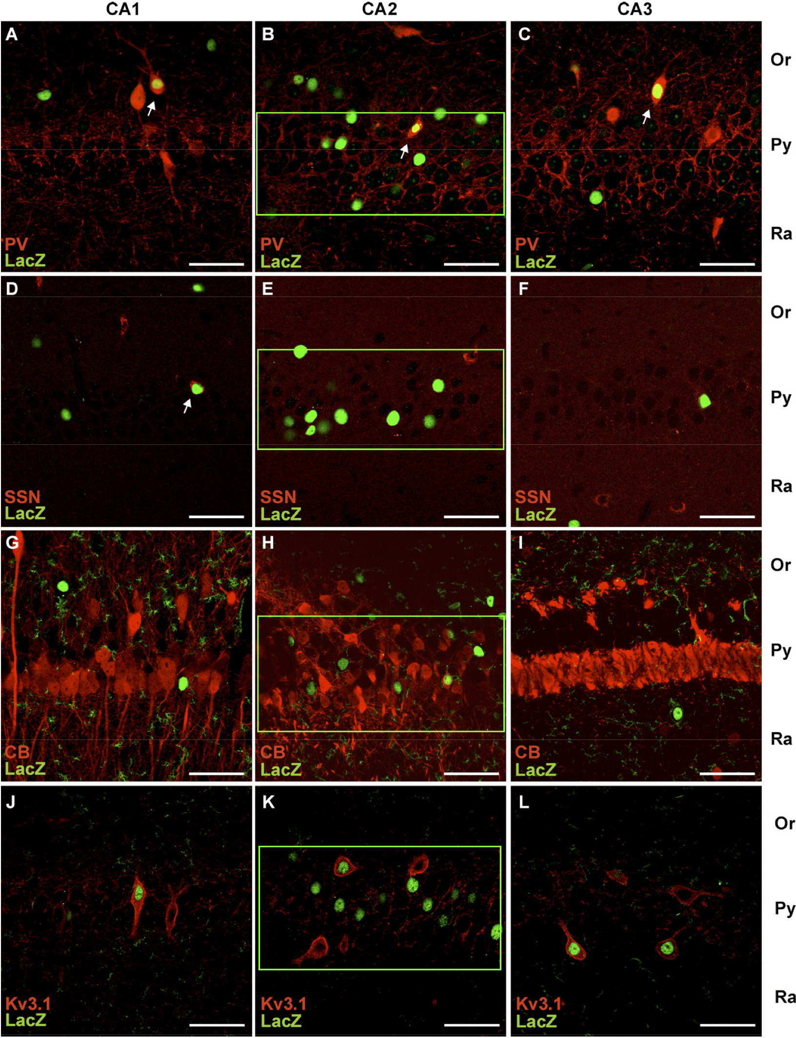
"Fig. 5. Co-immunofluorescence analyses with antibodies to beta-Gal (LacZ) and different interneuronal markers in hippocampal slices (100 [micro]m) of adult mice. (A - C) beta-Gal- and parvalbumin-positive signals were often colocalized within the areas CA1 (A) and CA3 (C) of the hippocampus. (D, E, F) Some somatostatin-positive interneurons, especially in CA1, demonstrated Cx30.2/LacZ expression. (G - I) Calbindin-positive interneurons in general did not show Cx30.2/LacZ expression. (J - L) Kv3.1-positive interneurons of the hippocampus were found to be beta-Gal-positive."
 Browse
 Search
 Help
 negative