All of the evidence provided on Hippocampome.org are quotes from scientific texts.
However, because quoted passages may be difficult to understand in isolation, contextual information and expanded abbreviations set in square brackets have been added for clarity.
Animal:
Protocol:
Expression:
Show:
EXPRESSION: positive
ANIMAL: mouse
PROTOCOL: mRNA
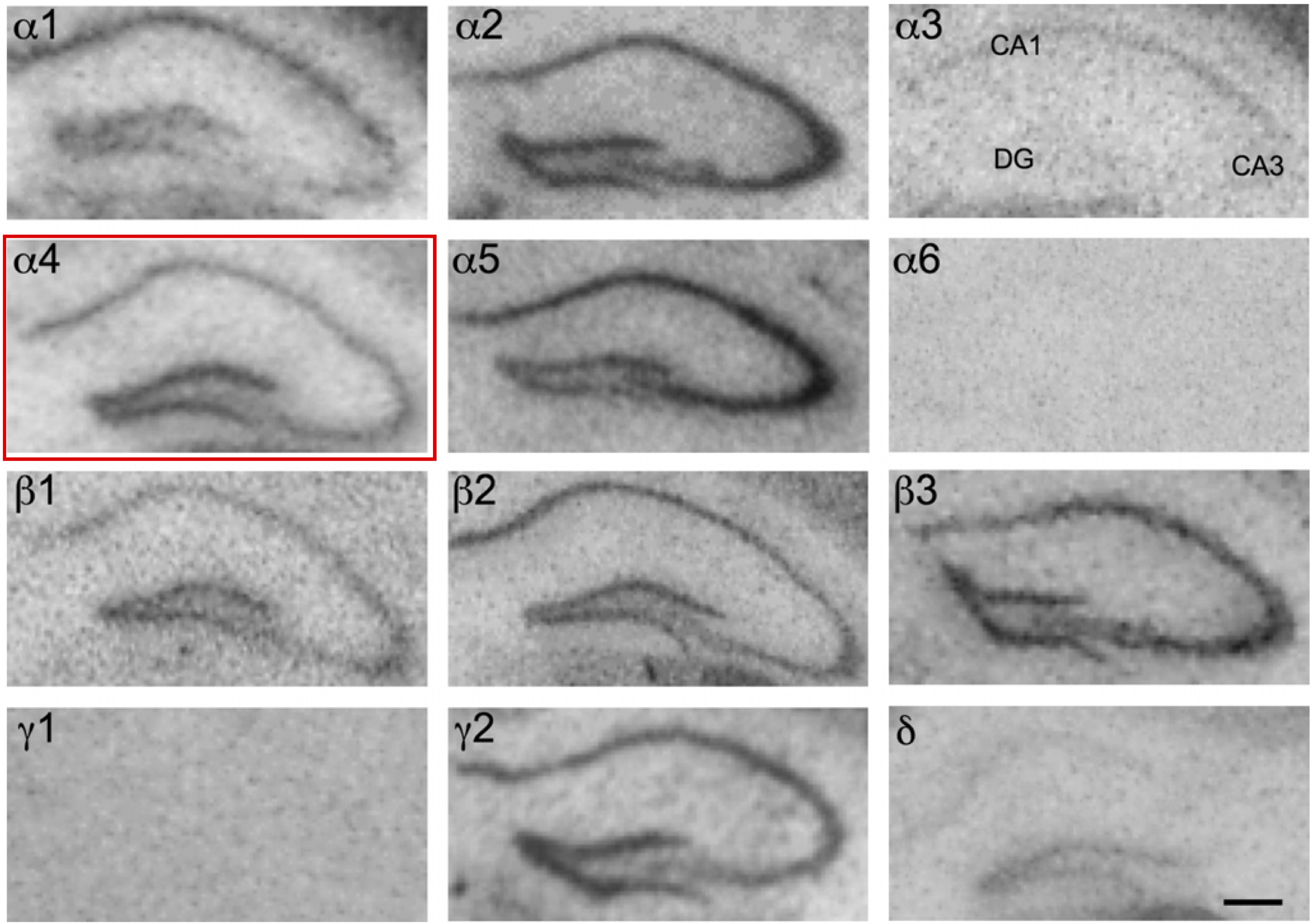
Page location: p353, Fig. 7
Linking PMID: 23337532 Linking Quote: Principal neurons identified by the presence of their somata in the principal cell layer, which is densely packed with and nearly homogeneous for the somata of the subregion's principal neurons (Czeh et al., 2013, Acta Biologica Hungarica).
"Fig. 7. Distribution of the GABA A receptor subunit mRNAs (a1-a6, b1-b3, c1, c2 and d) in the dorsal hippocampus (coronal sections of the mouse
brain approximately Bregma 1.82 mm, according to Franklin and Paxinos, 2007). In situ hybridization signals were restricted to the pyramidal cell
layer of cornu ammonis CA1 to CA3, the granule cell layer and the hilus of dentate gyrus. Note the strong signals of a2-, a5- and b3-subunits
throughout the hippocampus. a1-subunit mRNA labeling was weak in the CA3 area in comparison to a strong label in CA1 and dentate gyrus. a3-
subunit mRNA was weakly expressed and restricted to CA1, whereas d-subunit mRNA was almost exclusively restricted to the dentate gyrus."
 Browse
 Search
 Help
 positive