All of the evidence provided on Hippocampome.org are quotes from scientific texts.
However, because quoted passages may be difficult to understand in isolation, contextual information and expanded abbreviations set in square brackets have been added for clarity.
Animal:
Protocol:
Expression:
Show:
EXPRESSION: positive
ANIMAL: mouse
PROTOCOL: immunohistochemistry
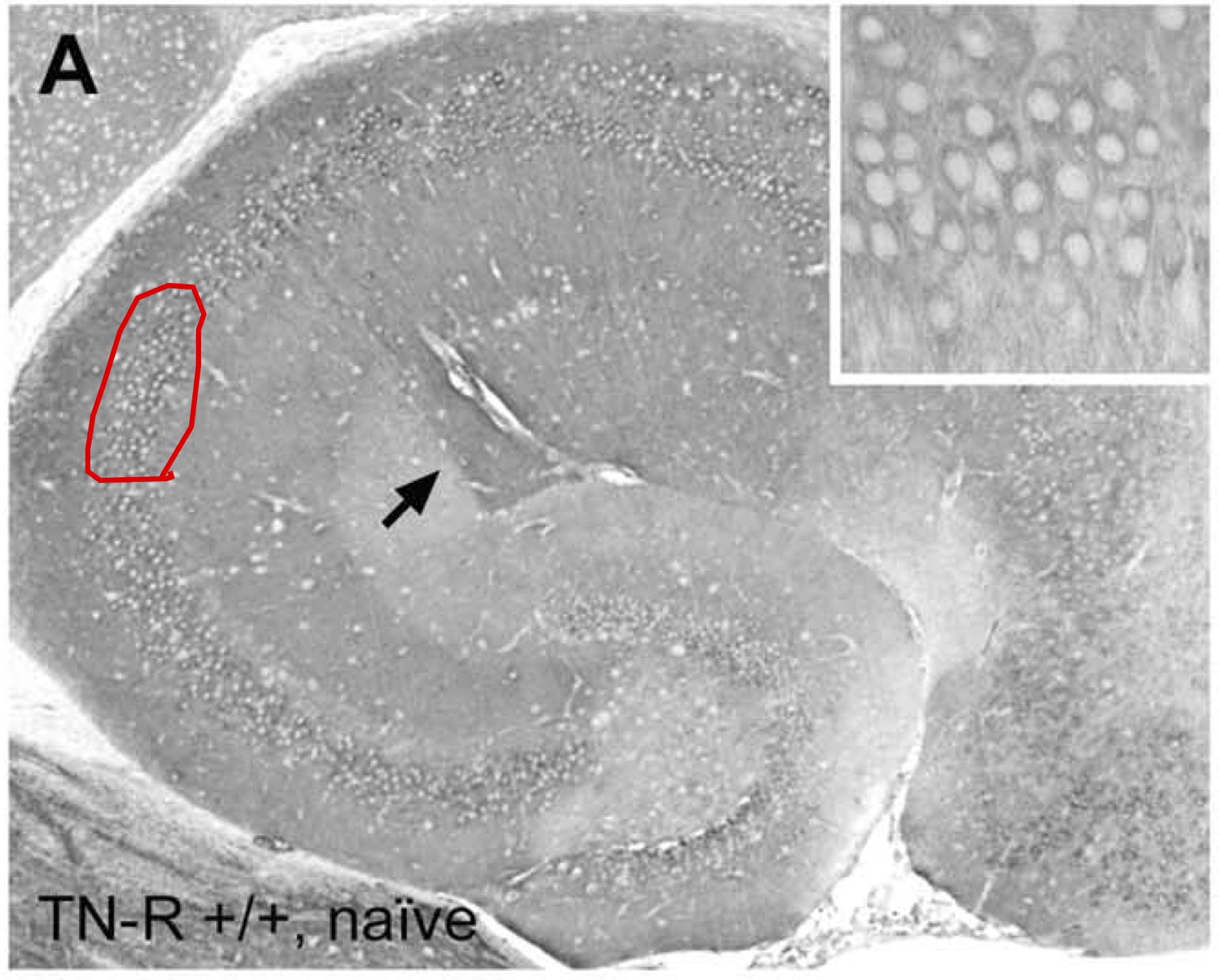
Page location: p847, Fig. 4
Linking PMID: 15026125 Linking Quote: Principal neurons identified by the presence of their somata in the principal cell layer, which is densely packed with and nearly homogeneous for the somata of the subregion's principal neurons (Czeh et al., 2013, Acta Biologica Hungarica).
"Fig. 4. Patterns of hippocampal calcineurin immunoreactivity. (A, B) Hippocampus of a naive wild-type (A) and TN-R-deficient (B) animal, showing
calcineurin staining in cell bodies of dentate granule cells, hilar neurons and pyramidal cells, as well as in dendrites and neuropil. Interestingly, the
staining intensity of the stratum lacunosum-moleculare is higher in CA1 compared with CA2 and CA3, due to a strong labeling of dendritic profiles
within the CA1 stratum lacunosum-moleculare (arrows). The insets in A and B demonstrate a magnified view of the respective CA3 pyramidal cell
layer. (C, D) Hippocampus of a wild-type (C) and TN-R-deficient (D) animal 10 weeks after pilocarpine treatment, showing increased calcineurin
immunoreactivity in the cell bodies and proximal dendrites of CA3 pyramidal neurons (see insets). The insets in C and D demonstrate a magnified view
of the respective CA3 pyramidal cell layer. The arrowheads point to examples of proximal dendrites with enhanced calcineurin immunoreactivity"
 Browse
 Search
 Help
 positive