All of the evidence provided on Hippocampome.org are quotes from scientific texts.
However, because quoted passages may be difficult to understand in isolation, contextual information and expanded abbreviations set in square brackets have been added for clarity.
Animal:
Protocol:
Expression:
Show:
Order:
EXPRESSION: positive
ANIMAL: mouse
PROTOCOL: immunohistochemistry
Page location: p225, right bottom
Linking PMID: 21516261 Linking Quote: Principal neurons identified by the presence of their somata in the principal cell layer, which is densely packed with and nearly homogeneous for the somata of the subregion's principal neurons (Czeh et al., 2013, Acta Biologica Hungarica).
"In hippocampus, Ng [neurogranin] and CaM [calmodulin] were colocalized in all principle neurons at the various subfields. Under the basal conditions, these proteins exhibited higher concentrations in soma than in dendrites with the exception of pyramidal neurons in subiculum complex, which exhibited a uniform distribution of both Ng and CaM in all cellular compartments (data not shown)."
EXPRESSION: positive
ANIMAL: mouse
PROTOCOL: immunohistochemistry
Page location: p225, Fig. 3
Linking PMID: 21516261 Linking Quote: Principal neurons identified by the presence of their somata in the principal cell layer, which is densely packed with and nearly homogeneous for the somata of the subregion's principal neurons (Czeh et al., 2013, Acta Biologica Hungarica).
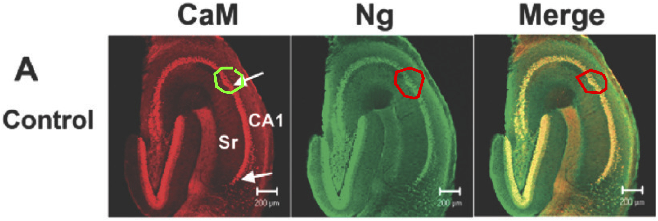
"Figure 3. Immunochemical staining of hippocampal slices sequentially treated with EGTA- and Ca(2+)-containing ACSF [artificial cerebrospinal fluid]. Fixed tissue sections (50 [micro]m) were doubly stained with antibodies against CaM [calmodulin] (Alexa 594) and Ng [neurogranin] (FITC) and the 5 confocal images of the whole sections of control (A), ..."
EXPRESSION: positive
ANIMAL: mouse
PROTOCOL: immunohistochemistry
Page location: p226, Fig. 4
Linking PMID: 21516261 Linking Quote: Principal neurons identified by the presence of their somata in the principal cell layer, which is densely packed with and nearly homogeneous for the somata of the subregion's principal neurons (Czeh et al., 2013, Acta Biologica Hungarica).
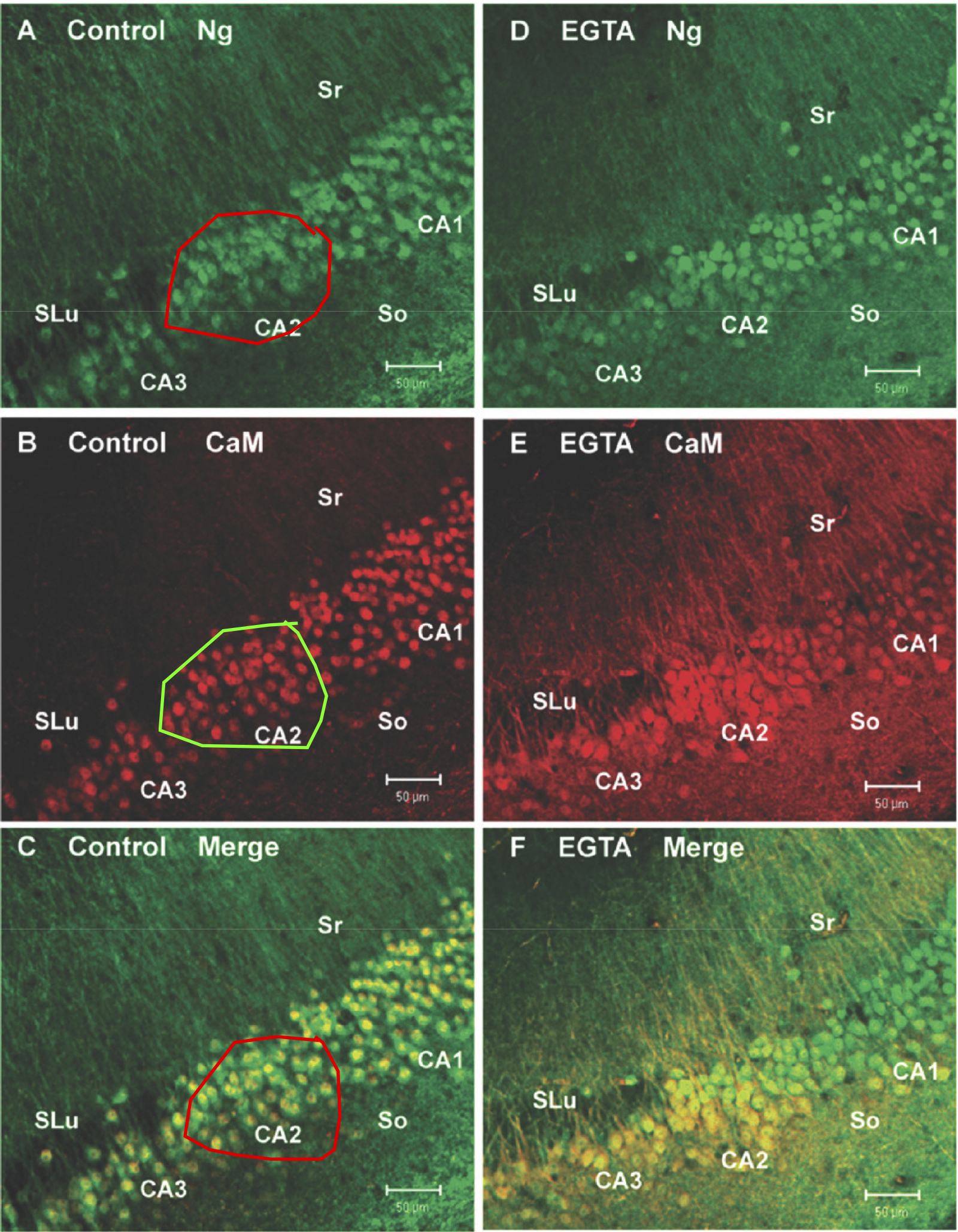
"Figure 4. Distinct responses among CA1, CA2, and CA3 neurons to treatment of EGTA/ACSF [EGTA-containing artificial cerebrospinal fluid]. Representative confocal images (10 x) show the control (A-C) and EGTA-treated (15 min) (D-F) samples doubly stained with antibodies against CaM [calmodulin] (Alexa 594) and Ng [neurogranin] (FITC). The CA3 region was identified by the appearance of stratum lucidem (SLu). Pyramidal neurons from CA2 and CA3 regions appeared slightly larger than those of the CA1. In the merged images of control (C), CaM was colocalized with Ng in the nucleus with a relatively low abundance in the cytoplasm and dendrites within stratum radiatum (Sr) and stratum oriens (So)."
 Browse
 Search
 Help
 positive