negative inference; confirmed by additional inferences
All of the evidence provided on Hippocampome.org are quotes from scientific texts.
However, because quoted passages may be difficult to understand in isolation, contextual information and expanded abbreviations set in square brackets have been added for clarity.
Animal:
Protocol:
Expression:
Show:
Order:
EXPRESSION: negative (inference)
ANIMAL: rat
PROTOCOL: immunohistochemistry
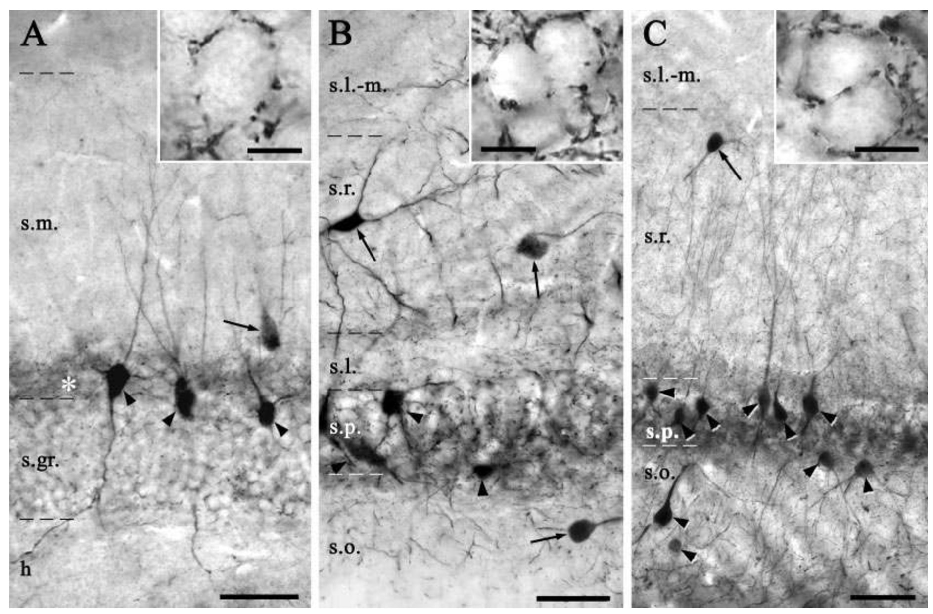
Page location: p463, Figure 2
Linking PMID: 15224983 Linking Quote: Principal neurons identified by the presence of their somata in the principal cell layer, which is densely packed with and nearly homogeneous for the somata of the subregion's principal neurons (Czeh et al., 2013, Acta Biologica Hungarica).
"FIGURE 2. Distribution of parvalbumin-immunoreactive (PV-IR) cells in the mouse hippocampus. A: In the dentate gyrus, PV-IR
cells could be found within or adjacent to stratum granulosum (arrowheads), occasionally, PV-containing interneurons could be observed in the hilus and stratum moleculare (arrow). Basket-like axon
terminals were found around the immunonegative granule cells (insert). In the inner third of stratum moleculare, a dense cloud of axon
staining could be seen (asterisk). B: In the CA3 region, PV-IR interneurons were present in all layers (arrows and arrowheads), except in
stratum lacunosum-moleculare. Most possessed radially oriented
dendrites (arrowheads), while the others in strata radiatum and oriens
had multipolar dendritic trees with horizontal branches (arrows).
Axon terminals could be seen in clusters around somata (insert), axon
initial segments and proximal basal dendrites of immunonegative
pyramidal cells. C: In the CA1 area, PV-IR interneurons were also
located in strata pyramidale and oriens (arrowheads). Most of them
showed the characteristic features of basket cells or chandelier cells.
Interneurons were rarely present in strata radiatum or lacunosum-moleculare (arrow). PV-IR axon terminals surround the somata and
axon initial segments of the pyramidal cells (insert) and their proximal dendrites."
Interpretation notes: Glutamatergic and GABAergic types in CA3 SLM are negative for PV
EXPRESSION: negative (inference)
ANIMAL: rat
PROTOCOL: immunohistochemistry
Page location: p971, left mid
"Parvalbumin-immunoreactive neurons were seen in strata oriens and pyramidale, but not in strata radiatum and lacunosum-moleculare of the CA1 and CA3, while parvalbumin-immunoreactive fibers were found in all strata, but more concentrated in the stratum pyramidale."
Interpretation notes: Glutamatergic and GABAergic types in CA3 SLM_SR are negative for PV
EXPRESSION: negative (inference)
ANIMAL: rat
PROTOCOL: immunohistochemistry
Page location: p 311, right bottom
"In the case of parvalbumin-VIP [vasoactive intestinal polypeptide] co-localization, only those VIP cells within or close to stratum pyramidale were examined, since parvalbumin cells are absent in other layers. Thus, all VIP-positive cells in the distal part of stratum radiatum or in stratum lacunosum-moleculare are considered parvalbumin-negative by their position. None of the 136 VIP-immunopositive cells near stratum pyramidale in CA1 and the 36 in CA3 showed parvalbumin immunofluorescence (Fig. 8E, F). In addition, 15 large multipolar VIPpositive basket cells were selected from different sections, but those were also negative for parvalbumin. This finding demonstrates that there is a basket cell type different from those containing parvalbumin but which is characterized by the coexistence of VIP and CCK [cholecystokinin]."
Interpretation notes: Glutamatergic and GABAergic types in CA3 SLM_SR are negative for PV
 Browse
 Search
 Help
 negative inference; confirmed by additional inferences