All of the evidence provided on Hippocampome.org are quotes from scientific texts.
However, because quoted passages may be difficult to understand in isolation, contextual information and expanded abbreviations set in square brackets have been added for clarity.
Animal:
Protocol:
Expression:
Show:
Order:
EXPRESSION: negative
ANIMAL: rat
PROTOCOL: mRNA
Page location: p285, Fig. 2
Linking PMID: 8170352 Linking Quote: Principal neurons identified by the presence of their somata in the principal cell layer, which is densely packed with and nearly homogeneous for the somata of the subregion's principal neurons (Czeh et al., 2013, Acta Biologica Hungarica).
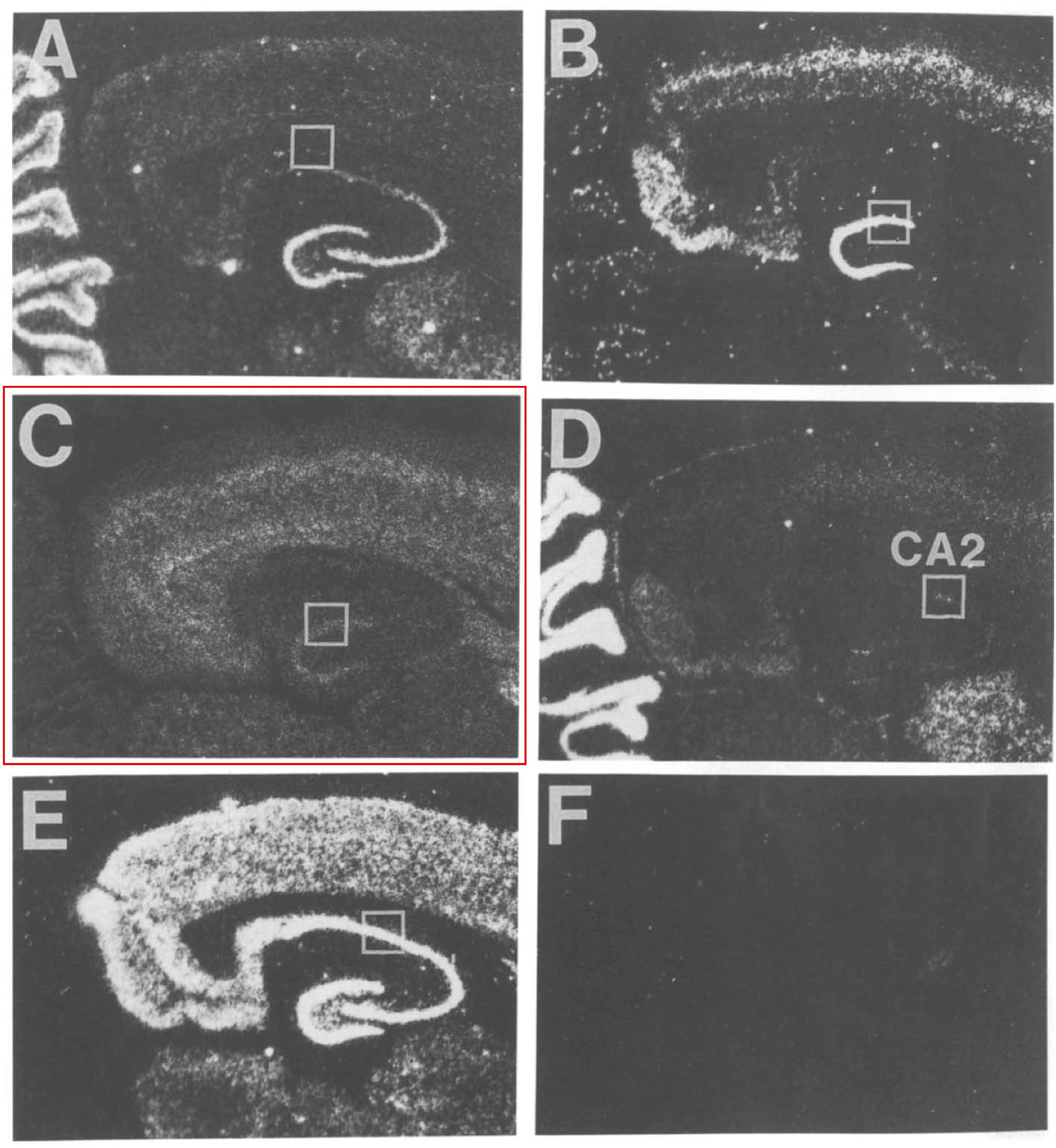
"Fig. 2. Dark-field images of hybridization signals of mGluR1 (A), mGluR2 (B), mGluR3 (C), mGluR4 (D), and mGluR5 (E) in the hippocampus
and entorhinal cortex. Positive labelling appears white. (F), mGluR5 with excess unlabelled probe."
EXPRESSION: negative
ANIMAL: rat
PROTOCOL: mRNA
Page location: p288, left middle
Linking PMID: 8170352 Linking Quote: Principal neurons identified by the presence of their somata in the principal cell layer, which is densely packed with and nearly homogeneous for the somata of the subregion's principal neurons (Czeh et al., 2013, Acta Biologica Hungarica).
"Within the hippocampus, mGluR3
labelling was most intense in the granular layer of the
dentate gyrus where both neuronal cell bodies and
small, densely-stained cells with scant cytoplasm, most
likely representative of glia, expressed this receptor
(Fig. 3C). The mGluR3 labelling in the pyramidal layer
of CA3 was at the same level as the labelling in the
adjacent white matter areas. However, the labelling in
the pyramidal layer of CA1 and in the subiculum was
significantly greater than the white matter areas."
 Browse
 Search
 Help
 negative