All of the evidence provided on Hippocampome.org are quotes from scientific texts.
However, because quoted passages may be difficult to understand in isolation, contextual information and expanded abbreviations set in square brackets have been added for clarity.
Animal:
Protocol:
Expression:
Show:
Order:
EXPRESSION: negative
ANIMAL: mouse
PROTOCOL: immunohistochemistry
Page location: p699, Fig. 3
Linking PMID: 21867885 Linking Quote: Principal neurons identified by the presence of their somata in the principal cell layer, which is densely packed with and nearly homogeneous for the somata of the subregion's principal neurons (Czeh et al., 2013, Acta Biologica Hungarica).
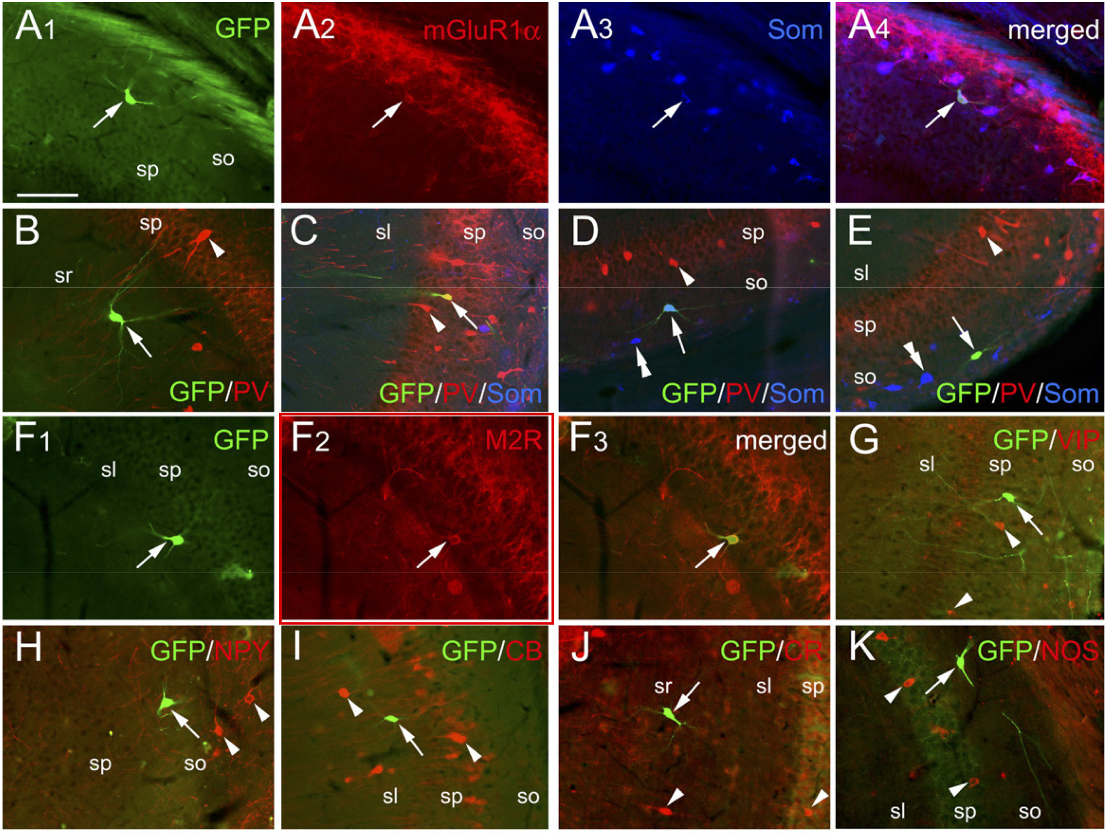
"Figure 3. SOM and mGluR1a Are Preferentially Expressed in EGins [early-generated interneurons] in the Adult Hippocampus
All images are from the CA3 area. All GFP-positive neurons correspond to EGins labeled using tamoxifen-treated Dlx1/2
CreERTM;RCE:LoxP mice (induction: E7.5).
(A) A GFP-positive EGin (arrow) coexpressing mGluR1a and SOM in the CA3 stratum oriens.
(B-E) Generally, EGins do not express PV (arrows in B, D, E) even though very occasional PV positive cells can be seen (arrow in C). GFP-positive cells never
coexpress PV and SOM (arrows in C-E) but can be observed next to cells that are positive for PV or SOM (single or double arrowheads). Note that the GFP cell in D
(arrow) is colabeled for SOM.
(F) M2R labeling in a GFP-positive cell (arrows).
(G-K) In the vast majority of fate-mapped GFP-positive cells (arrows), neither immunoreactivity (arrowheads) for VIP, nor for NPY, CB, CR, or NOS is detected."
EXPRESSION: negative
ANIMAL: rat
PROTOCOL: immunohistochemistry
Page location: p358, left bot
Linking PMID: 9466448 Linking Quote: Principal neurons identified by the presence of their somata in the principal cell layer, which is densely packed with and nearly homogeneous for the somata of the subregion's principal neurons (Czeh et al., 2013, Acta Biologica Hungarica).
"In the CA3 region, m2[muscarinic type 2 receptor]-IR [immunoreactive] neurons were located mostly in stratum oriens and had a multipolar morphology with three to four primary dendrites (Fig. 1D)."
EXPRESSION: negative
ANIMAL: rat
PROTOCOL: immunohistochemistry
Page location: p358, right mid
Linking PMID: 9466448 Linking Quote: Principal neurons identified by the presence of their somata in the principal cell layer, which is densely packed with and nearly homogeneous for the somata of the subregion's principal neurons (Czeh et al., 2013, Acta Biologica Hungarica).
"The most striking axonal staining for m2 [muscarinic type 2 receptor] was present in the perisomatic region of pyramidal cells. The dense plexus formed by fine fibres and large numbers of boutons surrounded immunonegative perikarya in a basket-like manner and outlined axon initial segments at the stratum pyramidale/oriens border (Fig. 1F). The immunoreactivity was stronger in the CA3 subfeld compared to CA1."
EXPRESSION: negative
ANIMAL: rat
PROTOCOL: immunohistochemistry
Page location: p362, right mid
Linking PMID: 9466448 Linking Quote: Principal neurons identified by the presence of their somata in the principal cell layer, which is densely packed with and nearly homogeneous for the somata of the subregion's principal neurons (Czeh et al., 2013, Acta Biologica Hungarica).
"The examination of GABA [gamma-aminobutyric acid] content showed that the vast majority (94%) of m2[muscarinic type 2 receptor]-IR[immunoreactive] cells [in CA1 and CA3] contained this marker (101 out of 107; Fig. 5A-D). The GABA-negative m2-IR neurons were found in stratum oriens and stratum radiatum of the CA3 region (3-3 neurons in both layers)."
EXPRESSION: negative
ANIMAL: rat
PROTOCOL: immunohistochemistry
Page location: p4081, right top
Linking PMID: 7751967 Linking Quote: Principal neurons identified by the presence of their somata in the principal cell layer, which is densely packed with and nearly homogeneous for the somata of the subregion's principal neurons (Czeh et al., 2013, Acta Biologica Hungarica).
"The pyramidal neuron layer [of CA1 and CA3] displayed a dense plexus of fine fibers and puncta outlining the perikarya, in basket-like formations, with the density of [muscarinic type 2 receptor] immunoreactivity greater in CA3 than in CA1. However, the pyramidal neurons themselves showed little or no immunoreactivity in the soma (Fig. 3)."
EXPRESSION: negative
ANIMAL: rat
PROTOCOL: immunohistochemistry
Page location: p4080, Fig. 2
Linking PMID: 7751967 Linking Quote: Principal neurons identified by the presence of their somata in the principal cell layer, which is densely packed with and nearly homogeneous for the somata of the subregion's principal neurons (Czeh et al., 2013, Acta Biologica Hungarica).
"Figure 2. Laminar distributions of m1-m4 immunoreactivity in CA1 and dentate gyrus. Each panel shows a similar column from coronal sections of dorsal hippocampus processed for Nissl (far left) or the muscarinic receptor immunoreactivities."
 Browse
 Search
 Help
 negative