All of the evidence provided on Hippocampome.org are quotes from scientific texts.
However, because quoted passages may be difficult to understand in isolation, contextual information and expanded abbreviations set in square brackets have been added for clarity.
Animal:
Protocol:
Expression:
Show:
Order:
EXPRESSION: positive
ANIMAL: mouse
PROTOCOL: immunohistochemistry
Page location: p497, Fig. 3
Linking PMID: 18620078 Linking Quote: Principal neurons identified by the presence of their somata in the principal cell layer, which is densely packed with and nearly homogeneous for the somata of the subregion's principal neurons (Czeh et al., 2013, Acta Biologica Hungarica).
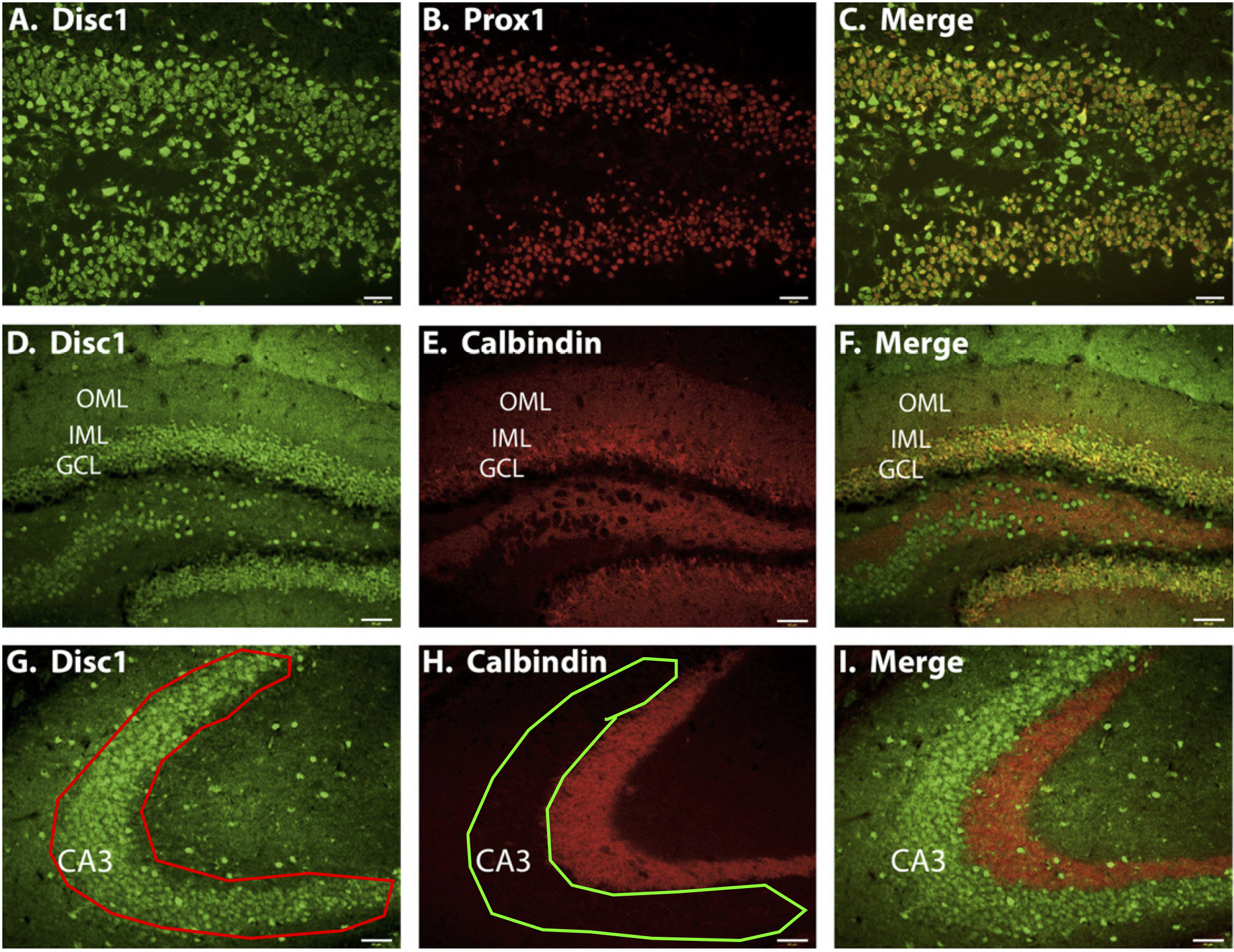
"Fig. 3. Disc1 expression in dentate gyrus granule cells. Disc1 is expressed in mature granule cells of the dentate gyrus as is shown by colocalization with the granule cell marker Prox1 (A-C; P7) and the mature granule cell marker calbindin (D-F; 6 weeks). Disc1 immunoreactivity is not detected with calbindin in the granule cell mossy fibers (G-I)."
EXPRESSION: positive
ANIMAL: mouse
PROTOCOL: immunohistochemistry
Page location: p498, left bottom
Linking PMID: 18620078 Linking Quote: Principal neurons identified by the presence of their somata in the principal cell layer, which is densely packed with and nearly homogeneous for the somata of the subregion's principal neurons (Czeh et al., 2013, Acta Biologica Hungarica).
"An examination of Disc1 subcellular localization in both CA3 pyramidal neurons and dentate gyrus granule cells reveals a strong expression pattern in discrete regions surrounding the nucleus (Fig. 6A-C). These data are consistent with previous studies which have revealed Disc1 localization in areas including perinuclear regions and the centrosome (Morris et al., 2003; James et al., 2004; Miyoshi et al., 2004; Kirkpatrick et al., 2006). Notably, we also observe that Disc1 expression extends slightly into the apical processes of CA3 neurons. This expression pattern is more pronounced during early postnatal development than in adulthood (Fig. 6, compare P7 (B) with 14 weeks (C)), perhaps indicative of a role for Disc1 in the outgrowth of dendritic processes during development."
EXPRESSION: positive
ANIMAL: mouse
PROTOCOL: immunohistochemistry
Page location: p496, Fig. 2
Linking PMID: 18620078 Linking Quote: Principal neurons identified by the presence of their somata in the principal cell layer, which is densely packed with and nearly homogeneous for the somata of the subregion's principal neurons (Czeh et al., 2013, Acta Biologica Hungarica).
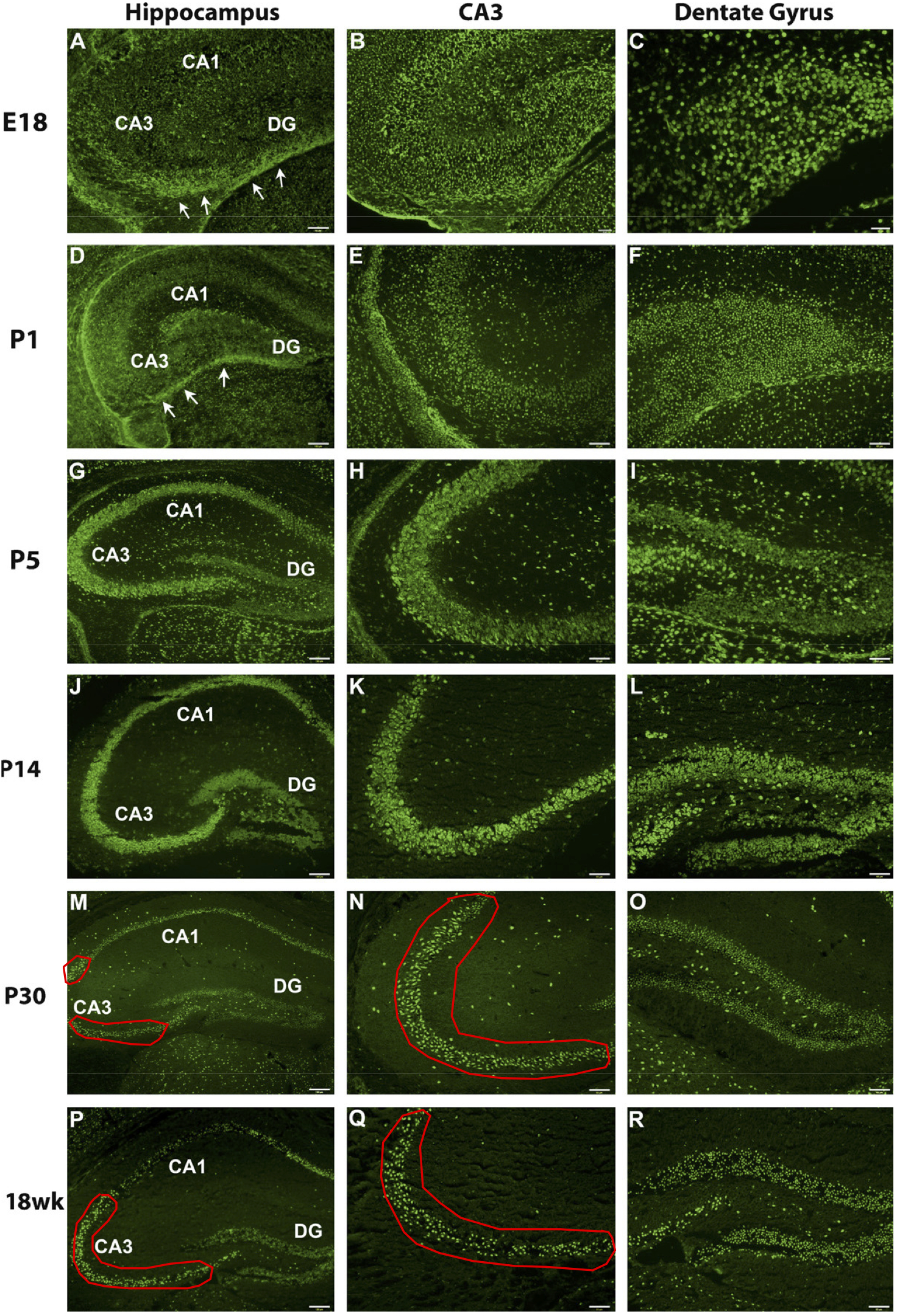
"Fig. 2. Disc1 hippocampal expression from E18-18 weeks. Disc1 is expressed in the hippocampus from embryonic development through adulthood. Both Disc1 (Mid) and (Cterm) antibodies were used to detect Disc1 expression in the mouse hippocampus. At E18 (A-C) and P1 (D-F), Disc1 is expressed throughout the hippocampus, which is still immature in its development. Disc1 is also detected in the stream of migrating granule cells and their precursors moving toward the dentate gyrus (arrows in A and D). From P5 (G-I) through P14 (J-L), Disc1's expression becomes more well-defined in Ammon�s horn and the dentate gyrus. . . ."
 Browse
 Search
 Help
 positive