All of the evidence provided on Hippocampome.org are quotes from scientific texts.
However, because quoted passages may be difficult to understand in isolation, contextual information and expanded abbreviations set in square brackets have been added for clarity.
Animal:
Protocol:
Expression:
Show:
Order:
EXPRESSION: positive, negative
ANIMAL: mouse
PROTOCOL: mRNA
Page location: p125, left bottom
Linking PMID: 22314660 Linking Quote: Principal neurons identified by the presence of their somata in the principal cell layer, which is densely packed with and nearly homogeneous for the somata of the subregion's principal neurons (Czeh et al., 2013, Acta Biologica Hungarica).
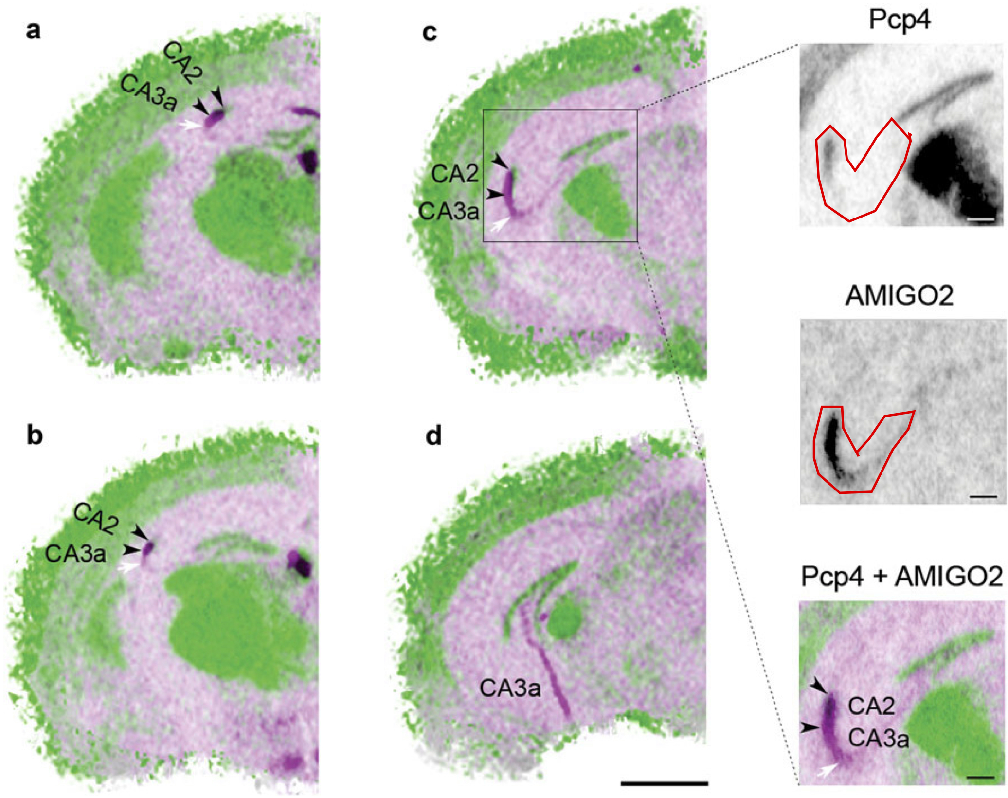
"A detailed comparison of AMIGO2 [amphoterin-induced gene and open reading frame 2]and Pcp4 [Purkinje cell protein 4] mRNA distribution patterns illustrates an overlap of expression in CA2. However, while Pcp4 mRNA expression was restricted to CA2, the AMIGO2 mRNA signal clearly extended further into the CA3 subfield, comprising CA3a in both the dorsal and the ventral horn (Figs. 2, 3; Ochiishi et al. 1999). In sagittal sections, the same distribution patterns could be discerned (Fig. 4). Pcp4 as well as AMIGO2 mRNA was clearly present in CA2 (Fig. 4, arrowheads), but only AMIGO2 mRNA was detected in the CA3a region."
EXPRESSION: positive, negative
ANIMAL: mouse
PROTOCOL: mRNA
Page location: p127, Fig. 2
Linking PMID: 22314660 Linking Quote: Principal neurons identified by the presence of their somata in the principal cell layer, which is densely packed with and nearly homogeneous for the somata of the subregion's principal neurons (Czeh et al., 2013, Acta Biologica Hungarica).
"Fig. 3 AMIGO2-Pcp4 [Purkinje cell protein 4] pseudo-colored overlays display the presence of AMIGO2 [amphoterin-induced gene and open reading frame 2] beyond CA2 into CA3a. AMIGO2 is represented in pink and Pcp4 in green on four different Bregma levels."
 Browse
 Search
 Help
 positive-negative: subtypes