negative inference; confirmed by additional inferences
All of the evidence provided on Hippocampome.org are quotes from scientific texts.
However, because quoted passages may be difficult to understand in isolation, contextual information and expanded abbreviations set in square brackets have been added for clarity.
Animal:
Protocol:
Expression:
Show:
Order:
EXPRESSION: negative (inference)
ANIMAL: rat
PROTOCOL: immunohistochemistry
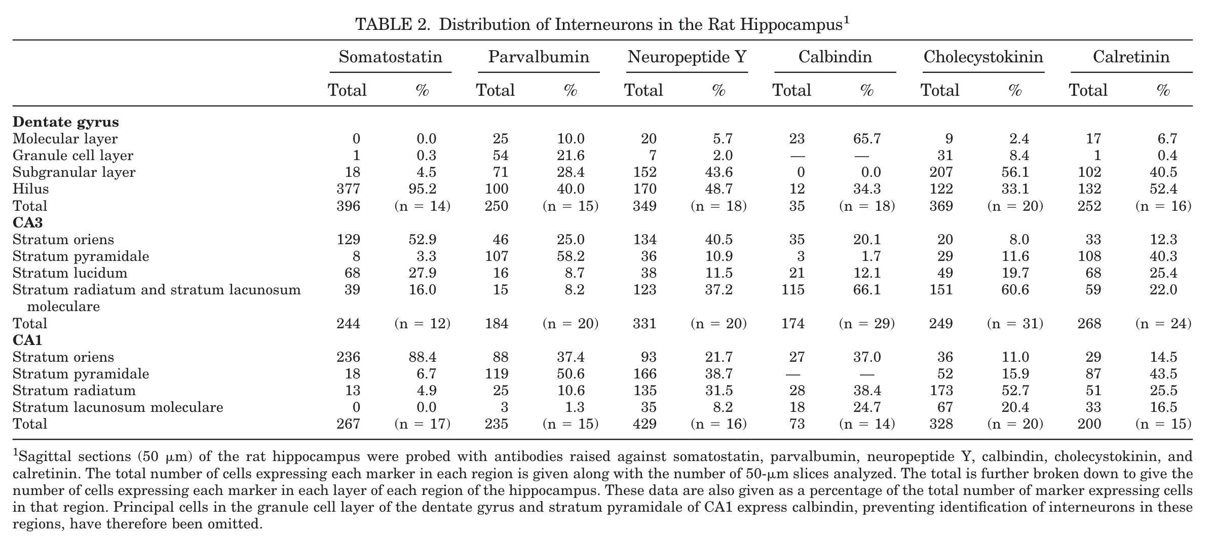
Page location: p554, table 2
Linking PMID: 18546278 Linking Quote: Principal neurons identified by the presence of their somata in the principal cell layer, which is densely packed with and nearly homogeneous for the somata of the subregion's principal neurons (Czeh et al., 2013, Acta Biologica Hungarica).
"TABLE 2. Distribution of Interneurons in the Rat Hippocampus Sagittal sections (50 micrometers) of the rat hippocampus were probed with antibodies raised against somatostatin, parvalbumin, neuropeptide Y, calbindin, cholecystokinin, and calretinin. The total number of cells expressing each marker in each region is given along with the number of 50-micrometer slices analyzed. The total is further broken down to give the number of cells expressing each marker in each layer of each region of the hippocampus. These data are also given as a percentage of the total number of marker expressing cells in that region. Principal cells in the granule cell layer of the dentate gyrus and stratum pyramidale of CA1 express calbindin, preventing identification of interneurons in these regions, have therefore been omitted."
Interpretation notes: Glutamatergic and GABAergic types in CA1 SLM are negative for SOM
EXPRESSION: negative (inference)
ANIMAL: rat
PROTOCOL: immunohistochemistry
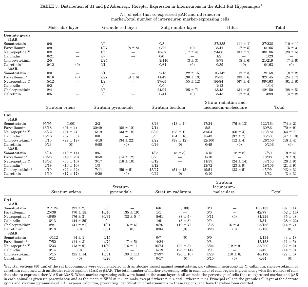
Page location: p555, table 3
"TABLE 3. Distribution of 1 and 2 Adrenergic Receptor Expression in Interneurons in the Adult Rat Hippocampus"
Interpretation notes: Glutamatergic and GABAergic types in CA1 SLM are negative for SOM
EXPRESSION: negative (inference)
ANIMAL: mouse
PROTOCOL: immunohistochemistry
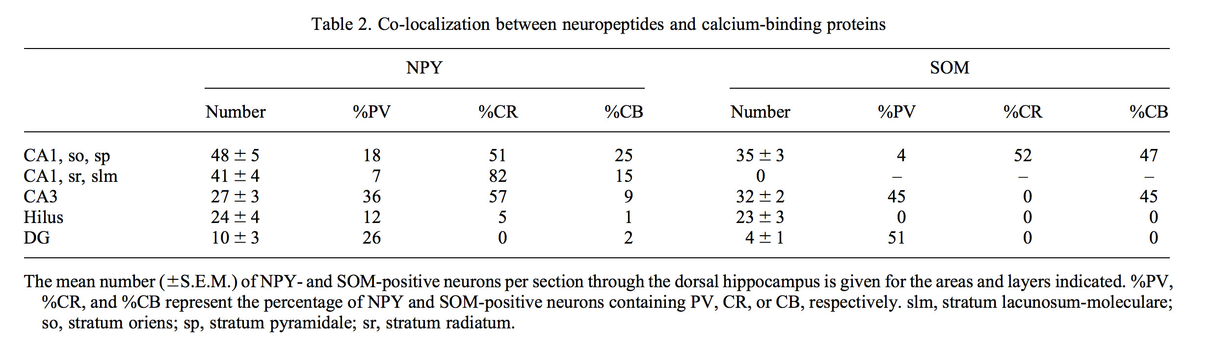
Page location: p49, Table 2
"Table 2. Co-localization between neuropeptides and calcium-binding proteins"
Interpretation notes: Glutamatergic and GABAergic types in CA1 SLM_SR are negative for SOM
EXPRESSION: negative (inference)
ANIMAL: rat
PROTOCOL: immunohistochemistry
Page location: p392, right bottom
"Hippocampus. SOM [somatostatin]-immunoreactive cell bodies are primarily
confined to stratum oriens in the CA1 region, with only a small number of cells in stratum pyramidale, and practically none in
strata radiatum and lacunosum-moleculare (Fig. 20). They are
more widespread in the CA3 region and frequently occur in strata
pyramidale, lucidum, and proximal radiatum."
Interpretation notes: Glutamatergic and GABAergic types in CA1 SLM_SR are negative for SOM
EXPRESSION: negative (inference)
ANIMAL: rat
PROTOCOL: immunohistochemistry
Page location: p775, right mid
"In these experiments, all the cells of the hippocampus immunoreactive for somatostatin were strongly immunoreactive for mGluR[metabotropic glutamate receptor]lalpha, and likewise the cells immunoreactive for mGluRla were also immunoreactive for somatostatin (Figures 2A and 2B)."
Interpretation notes: Glutamatergic and GABAergic mGluR1a- types in CA1 SLM_SR_SP_SO are probably SOM- by contrapositive logic
 Browse
 Search
 Help
 negative inference; confirmed by additional inferences