Sub CA1-Projecting Pyramidal
* indicates new v2.0 neuron type
Name Supertype (ID:Family:Type:Subregion)
Sub (e)331p-CA1_1110 CA1-Projecting Pyramidal S040405:Glutamatergic PC:Pyramidal:Sub

Name derivation
This is the only name in the cited literature. The modifer distinguishes it from the EC-targeting variant. Sub distinguishes these cells from pyramidal cells in other subregions.

Synonym(s)
subicular pyramidal cell
subicular pyramidal neuron

List of articles
Click here to view the list
Chang EH  (2012) Frontiers in Behavioral Neuroscience
Neurophysiological correlates of object recognition in the dorsal subiculum
Chang EH, Huerta PT.
Frontiers in Behavioral Neuroscience, 2012, 9 (46), Pages: 1 - 13
PMID: 22833721; DOI: 10.3389/fnbeh.2012.00046
Tags: phases
Chang EH  (2012) Frontiers in Behavioral Neuroscience
Neurophysiological correlates of object recognition in the dorsal subiculum
Chang EH, Huerta PT.
Frontiers in Behavioral Neuroscience, 2012, 9 (46), Pages: 1 - 13
PMID: 22833721; DOI: 10.3389/fnbeh.2012.00046
Tags: phases
Harris E  (2001) Brain Res
Propagation of synchronous epileptiform events from subiculum backward into area CA1 of rat brain slices.
Harris E, Stewart M.
Brain Res, 2001 Mar 23, 895 (2-Jan), Pages: 41 - 49
PMID: 11259758; DOI: 10.1016/S0006-8993(01)02023-6
Tags: morphology, connectivity, synapse probabilities

List of Hippocampome.org to NeuroMorpho.Org mappings
Click here to view the list of mappings
  • Ascoli
    • sub (fits the general description of "Subicular Pyramidal" cells)
  • MouseLight
  • Peng
  • Radley
  • Rice-Baylor
  • Schmitz
  • Sikdar
    • pyramid (fits the general description of "Subicular Pyramidal" cells)
  • Spruston
    • RS (fits the general description of "Subicular Pyramidal" cells)

  • Morphology
    Soma
    SUB:SP
    Axons
    CA1:SLM
    CA1:SR
    CA1:SP
    SUB:SM
    SUB:SP
    SUB:PL
    Dendrites
    SUB:SM
    SUB:SP

    Representative figure
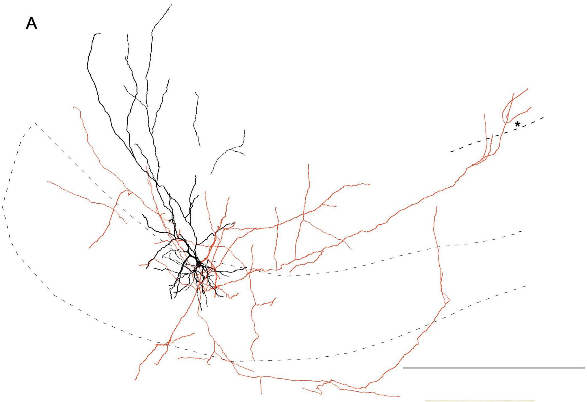
    Propagation of synchronous epileptiform events from subiculum backward into area CA1 of rat brain slices.
    Harris E, Stewart M
    Brain Res, 2001 Mar 23, 895 (2-Jan), pages: 41 - 49
    PMID: 11259758; DOI: 10.1016/S0006-8993(01)02023-6


    "Fig. 2. Neurolucida reconstruction of a neurobiotin-labeled subicular pyramidal neuron {Sub CA1-Projecting Pyramidal} to illustrate axon collaterals that reach the apical dendrites of CA1. Regular spiking neuron in rat subiculum has an axon collateral that ascends obliquely into the distal apical dendritic region of CA1 where it appears to have several potential synaptic contact points, evidenced by axonal extensions. The putative terminals at about the boundary between the stratum radiatum and the stratum lacunosum/moleculare. Another collateral runs just beneath the cell layer and has a branch that ascends through the cell layer in the transitional region between CA1 and subiculum. (A) Neurolucida reconstruction. The cell layer is outlined by a dashed line. A dashed line segment in CA1 marks the approximate boundary between stratum radiatum and stratum lacunosum/moleculare. The asterisk below the axon segment in CA1 corresponds to the asterisk location shown in B2. (B1) Low power view of the subiculum-CA1 border. The cell soma or proximal apical dendrites are not present in this section. This section contains the terminal axon in area CA1, shown at higher magnification in B2. (B2) High power view of axon terminals in the distal apical dendritic region of CA1. The visible axonal extensions (one marked by an asterisk) suggestive of synaptic contacts were present near the border of stratum radiatum with stratum lacunosum/moleculare. Scale bars: (A) 500 [micro]m; (B1) 200 [micro]m; (B2) 50 [micro]m."


    Molecular markers
    Positive
    CaM
    GABAa\alpha 2
    GABAa\alpha 3
    GABAa\alpha 4
    GABAa\alpha 5
    GABAa\beta 1
    GABAa\beta 2
    GABAa\beta 3
    GABAa\gamma 2
    Gaba-a-alpha
    mGluR5
    NG
    Negative
    5HT-3
    CB
    CB1
    CCK
    ENK
    ErbB4
    GABAa \delta
    GABAa\alpha 6 (confirmed by inference)
    NPY
    PV
    RLN
    SOM
    VIP
    Mixed expression
    nNOS (subtypes)

    Electrophysiological properties
    Key: [range] OR representative value±SD (measurements); Number of sources (total measurements): [min , max]

    Resting membrane potential (Vrest): -60 mV (1); 1 source
    Input resistance (Rin): [140.0, 160.0] MΩ (2); 1 source
    Threshold potential (Vthresh): 11 mV (1); 1 source
    Fast afterhyperpolaziring potential amplitude (Fast AHP): 8 mV (1); 1 source
    Action potential amplitude (APampl): 41 mV (1); 1 source
    Slow afterhyperpolarizing potential amplitude (Slow AHP): 2 mV (1); 1 source

    Notes
    Related classes: SUB (+)331-EC 000111 pyramidal neurons (active, need Giorgio approval) Harris (2001): Axonal reach into CA1:SP less than 200 microm; regular spiking. Finch (1983)(not yet cited): Single collateral reaching CA1 SO. Köhler (1985) (not yet cited): labeled fibers apparent in CA1 SM_SO. Witter email correspondence: "Menno's recommendation is that we separate the subicular pyramids into two distinct classes, one projecting to EC and the other to CA1. The logic is weak, but here goes the argument: there's is enough evidence to define 2 classes based on the input connectivity anyway. The more superficial pyramids (within PC but towards ML) have longer & thinner dendrites that receive input from L3 EC pyramidal cells, and these cells do NOT project to EC. In contrast, the deeper subicular pyramids (within PC but towards PL) do NOT receive EC L3 perforant path projections, and they DO project to EC (L5+6). The less clear cut assumption is that the former subicular pyramids form the (less abundant) back-projection to CA1, and the latter cells do not. The reasoning is that in ALL extra-hippocampal projections subicular neurons exclusively target ONE region and do not collateralize to multiple regions. Moreover, the two pyramidal subtypes also correspond to the bursting / regular firing phenotype. The evidence for the above is in Witter's paper in the subiculum special issue (2006?). He gave permission to quote him as "personal communication."

    Firing Pattern
    Images
    Image Missing
    Parameters View page
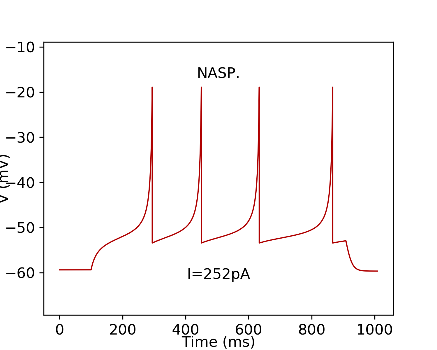
    NASP [Evidence] [Firing Pattern]
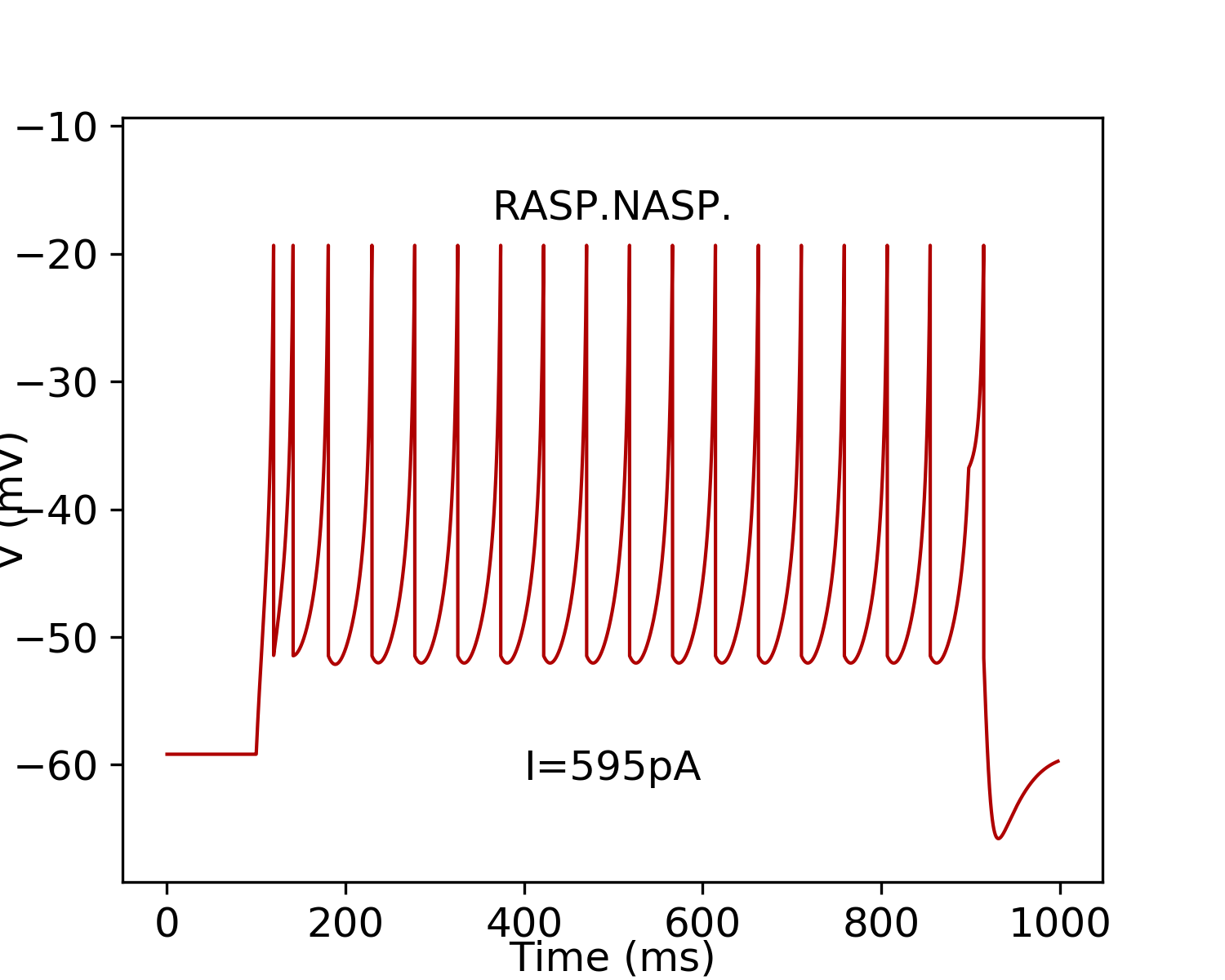
    RASP.ASP. [Evidence] [Firing Pattern]

    Izhikevich Model
    Parameters Single Compartment Downloads Simulate
    subtype: 1 k=4.64;a= 0;b= 25.16;d= 3;C= 724;Vr= -59.4;Vt= -45.35;Vpeak= -18.92;Vmin= -53.45; Simulate
    subtype: 2 k=2.47;a= 0.028;b= 25.94;d= 99;C= 372;Vr= -59.17;Vt= -44.5;Vpeak= -19.35;Vmin= -51.46; Simulate
    Images
    Image MissingImage Missing
    ModelDB Model
    Model Accession Number PubMed ID
    206365 27852771

    Sources of Input
    Known sources
    Excitatory or Inhibitory
    Sub Axo-axonic

    Potential sources
    Excitatory or Inhibitory
    DG Neurogliaform
    DG Wide-Arbor Neurogliaform*
    CA3c Pyramidal
    CA1 Pyramidal
    CA1 Deep Pyramidal
    CA1 Superficial Pyramidal
    CA1 Oriens-QuadA Projecting
    CA1 Perforant Path-Associated
    CA1 SO-SO Projecting*
    CA1 Trilaminar
    Sub CA1-Projecting Pyramidal
    Sub EC-Projecting Pyramidal
    Sub Pyramidale Multipolar Interneuron*
    Sub Recurrent Polymorphic-Moleculare*
    EC LI-II Pyramidal-Fan
    MEC LII Stellate
    EC LII-III Pyramidal-Tripolar
    EC LIII Small Pyramidal
    EC LIII Pyramidal
    EC LIV-VI Deep Multipolar Principal
    MEC LV Pyramidal

    Potential sources known to be avoided
    Excitatory or Inhibitory
    none known

    Targets of Output
    Known targets
    Excitatory or Inhibitory
    CA1 Pyramidal

    Potential targets
    Excitatory or Inhibitory
    CA2 Wide-Arbor Basket
    CA1 Deep Pyramidal
    CA1 Superficial Pyramidal
    CA1 Cajal-Retzius
    CA1 Local-Projecting Cajal-Retzius*
    CA1 Radiatum Giant
    CA1 Axo-axonic
    CA1 Back-Projection
    CA1 Basket
    CA1 R-P Basket*
    CA1 Trilayer Basket*
    CA1 Vertical Basket*
    CA1 Basket CCK+
    CA1 Trilayer Basket CCK+*
    CA1 Bistratified
    CA1 RO-receiving CA3-targeting*
    CA1 Hippocampo-subicular Projecting ENK+
    CA1 Ivy
    CA1 Interneuron Specific LM-R
    CA1 Interneuron Specific LMO-O
    CA1 LMR
    CA1 LMR-LM*
    CA1 Interneuron Specific LMR-O*
    CA1 LMR-R*
    CA1 Interneuron Specific LMR-R
    CA1 LMR Projecting
    CA1 Neurogliaform
    CA1 Neurogliaform Projecting
    CA1 Interneuron Specific O-Targeting QuadD
    CA1 Oriens/Alveus
    CA1 OR-LM
    CA1 Perforant Path-Associated
    CA1 Perforant Path-Associated QuadD
    CA1 QuadD-RP*
    CA1 Quadrilaminar
    CA1 Interneuron Specific R-O
    CA1 R-Receiving Apical-Targeting
    CA1 R-RO*
    CA1 R-RP*
    CA1 Radiatum
    CA1 Interneuron Specific RO-O
    CA1 Interneuron Specific RO-R*
    CA1 RO-RP*
    CA1 RP-RPO*
    CA1 Schaffer Collateral-Associated
    CA1 Schaffer Collateral-Correlated*
    CA1 Schaffer Collateral-Receiving R-Targeting
    CA1 Radial Trilaminar
    Sub CA1-Projecting Pyramidal
    Sub EC-Projecting Pyramidal
    Sub Axo-axonic
    Sub Pyramidale Multipolar Interneuron*
    Sub Wide-arbor Polymorphic*
    Sub Recurrent Polymorphic-Moleculare*

    Potential targets known to be avoided
    Excitatory or Inhibitory
    none known

    Oscillation Phase Locking
    Category
    Value
    Theta (deg):105.00
    Theta range:[105.00, 105.00]
    Theta measurements:1
    Theta representative selection:   mice, adult, female,
    tetrode, ketamine, xylazine, and acepromazine
    In vivo firing rate (Hz):1.24
    Firing rate range:[1.24, 1.24]
    Firing rate measurements:1
    Firing rate representative selection:   mice, adult, female,
    tetrode, ketamine, xylazine, and acepromazine