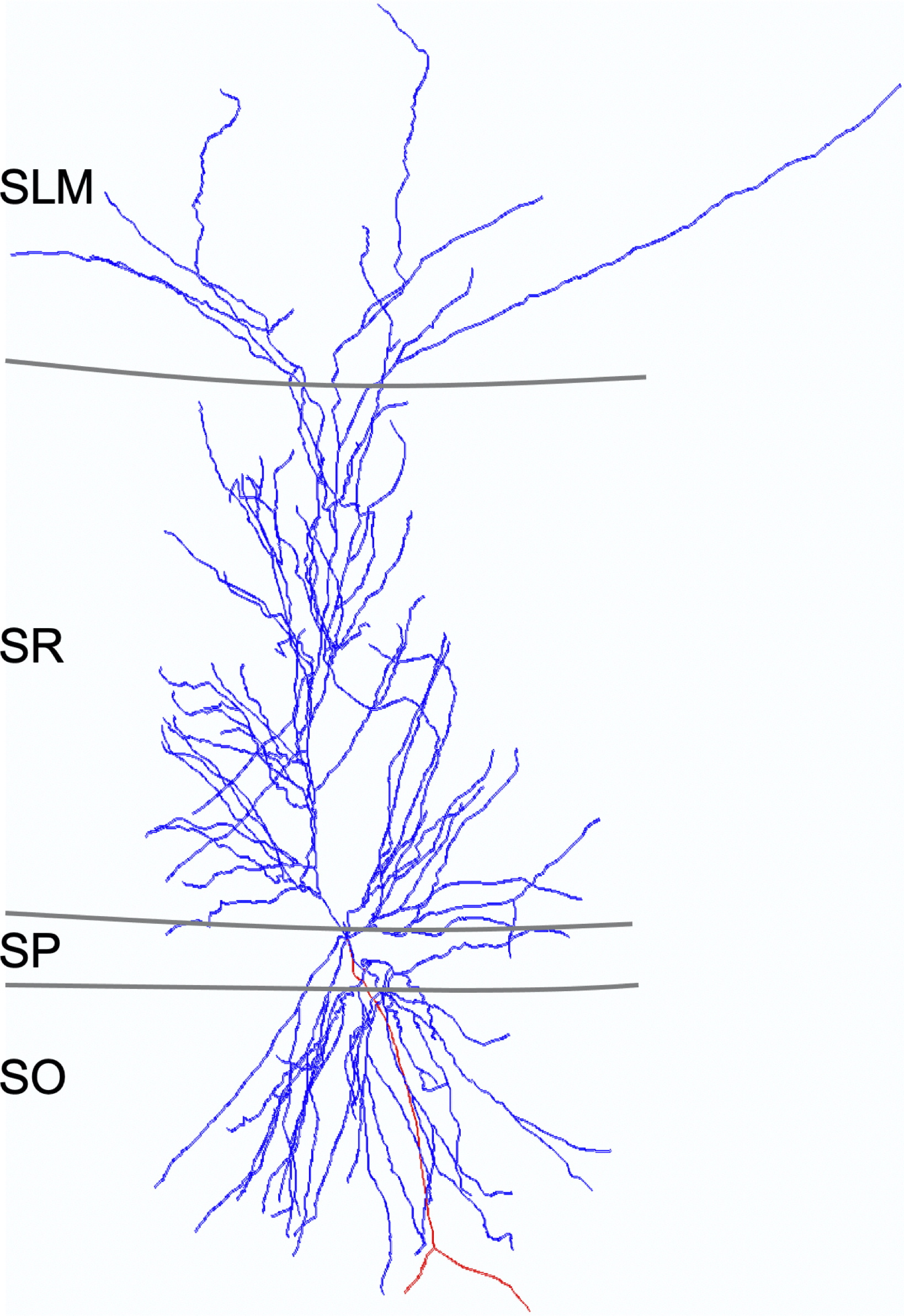
CA1 Superficial Pyramidal
* indicates new v2.0 neuron type
Name Supertype (ID:Family:Type:Subregion)
CA1 (e)2223p-Sub_111-EC_000011 Superficial Pyramidal S040404:Glutamatergic PC:Pyramidal:CA1

Name derivation
This is a canonical principal cell of the main hippocampal circuit. CA1 distinguishes these cells from pyramidal cells in other subregions. The phrase "Superficial" denotes the fact that these cells tend to be found more toward the top of SP.

Synonym(s)

List of articles
Click here to view the list
Amaral D  (2007) The Hippocampus Book
Hippocampal Neuroanatomy
Amaral D, Lavenex P.
The Hippocampus Book, 2007, Pages: 35 - 112
ISBN: 9780195100273
Tags: morphology, connectivity, synapse probabilities
Arszovszki A  (2014) Front Neuroanat
Three axonal projection routes of individual pyramidal cells in the ventral CA1 hippocampus
Arszovszki A, Borhegyi Z, Klausberger T.
Front Neuroanat, 2014, 8 (53), Pages: 1 - 11
PMID: 25009471; DOI: 10.3389/fnana.2014.00053
Tags: morphology
Bartos M  (2010) Hippocampal Microcircuits: A Computational Modeler's Resource Book
Fast and Slow GABAergic Transmission in Hippocampal Circuits
Bartos M, Sauer JF, Vida I, Kulik A.
Hippocampal Microcircuits: A Computational Modeler's Resource Book, 2010, Pages: 129 - 162
ISBN: 9781441909954
Tags: morphology, connectivity, synapse probabilities
Bernard C  (1994) Hippocampus
Model of local connectivity patterns in CA3 and CA1 areas of the hippocampus.
Bernard C, Wheal HV.
Hippocampus, 1994 Oct, 4 (5), Pages: 497 - 529
PMID: 7889123; DOI: 10.1002/hipo.450040502
Tags: morphology, connectivity, synapse probabilities
Freund TF  (1996) Hippocampus
Interneurons of the hippocampus.
Freund TF, Buzsaki G.
Hippocampus, 1996, 6 (4), Pages: 347 - 470
PMID: 8915675; DOI: 10.1002/(SICI)1098-1063(1996)6:4<347::AID-HIPO1>3.0.CO;2-I
Tags: morphology, connectivity, synapse probabilities
Fuentealba P  (2010) J Neurosci
Expression of COUP-TFII nuclear receptor in restricted GABAergic neuronal populations in the adult rat hippocampus.
Fuentealba P, Klausberger T, Karayannis T, Suen WY, Huck J, Tomioka R, Rockland K, Capogna M, Studer M, Morales M, Somogyi P.
J Neurosci, 2010 Feb 3, 30 (5), Pages: 1595 - 1609
PMID: 20130170; DOI: 10.1523/JNEUROSCI.4199-09.2010
Tags: morphology, connectivity, phases, synapse probabilities
Gloveli T  (2010) Hippocampal Microcircuits: A Computational Modeler's Resource Book
Neuronal Activity Patterns During Hippocampal Network Oscillations In Vitro
Gloveli T, Kopell N, Dugladze T.
Hippocampal Microcircuits: A Computational Modeler's Resource Book, 2010, Pages: 247 - 276
ISBN: 9781441909954
Tags: morphology, connectivity, synapse probabilities
Klausberger T  (2008) Science
Neuronal diversity and temporal dynamics: the unity of hippocampal circuit operations.
Klausberger T, Somogyi P.
Science, 2008 Jul 4, 321 (5885), Pages: 53 - 57
PMID: 18599766; DOI: 10.1126/science.1149381
Tags: morphology, connectivity, synapse probabilities
Klausberger T  (2009) Eur J Neurosci
GABAergic interneurons targeting dendrites of pyramidal cells in the CA1 area of the hippocampus.
Klausberger T.
Eur J Neurosci, 2009 Sep, 30 (6), Pages: 947 - 957
PMID: 19735288; DOI: 10.1111/j.1460-9568.2009.06913.x
Tags: morphology, connectivity, synapse probabilities
Lapray D  (2012) Nat Neurosci
Behavior-dependent specialization of identified hippocampal interneurons.
Lapray D, Lasztoczi B, Lagler M, Viney TJ, Katona L, Valenti O, Hartwich K, Borhegyi Z, Somogyi P, Klausberger T.
Nat Neurosci, 2012 Sep, 15 (9), Pages: 1265 - 1271
PMID: 22864613; DOI: 10.1038/nn.3176
Tags: marker, phases, synapse probabilities
Mizuseki K  (2011) Nat Neurosci.
Hippocampal CA1 pyramidal cells form functionally distinct sublayers.
Mizuseki K, Diba K, Pastalkova E, Buzsaki G.
Nat Neurosci., 2011, Pages: 1174 - 1181
PMID: 21822270
Tags: phases
Naber PA  (2001) Hippocampus
Reciprocal connections between the entorhinal cortex and hippocampal fields CA1 and the subiculum are in register with the projections from CA1 to the subiculum.
Naber PA, Lopes da Silva FH, Witter MP.
Hippocampus, 2001, 11 (2), Pages: 99 - 104
PMID: 11345131; DOI: 10.1002/hipo.1028
Tags: morphology
Navas-Olive A  (2020) Nature Communications
Multimodal determinants of phase-locked dynamics across deep-superficial hippocampal sublayers during theta oscillations
Navas-Olive A, Valero M, Jurado-Parras T, de Salas-Quiroga A, Averkin RG, Gambino G, Cid E, de la Prida LM.
Nature Communications, 2020, Pages: 2217 -
PMID: 32371879
Tags: phases
Oliva A  (2016) Hippocampus
Spatial Coding and Physiological Properties of Hippocampal Neurons in the Cornu Ammonis Subregions
Oliva A, Fernandez-Ruiz A, Buzsaki G, Berenyi A.
Hippocampus, 2016, Pages: 1593 - 1607
PMID: 27650887
Tags: phases
Pawelzik H  (2002) J Comp Neurol
Physiological and morphological diversity of immunocytochemically defined parvalbumin- and cholecystokinin-positive interneurones in CA1 of the adult rat hippocampus.
Pawelzik H, Hughes DI, Thomson AM.
J Comp Neurol, 2002 Feb 18, 443 (4), Pages: 346 - 367
PMID: 11807843; DOI: 10.1002/cne.10118
Tags: morphology, connectivity, synapse probabilities
Price CJ  (2005) J Neurosci
Neurogliaform neurons form a novel inhibitory network in the hippocampal CA1 area.
Price CJ, Cauli B, Kovacs ER, Kulik A, Lambolez B, Shigemoto R, Capogna M.
J Neurosci, 2005 Jul 20, 25 (29), Pages: 6775 - 6786
PMID: 16033887; DOI: 10.1523/JNEUROSCI.1135-05.2005
Tags: morphology, connectivity, synapse probabilities
Sanchez-Aguilera A  (2021) PLoS Biology
An update to Hippocampome.org by integrating single-cell phenotypes with circuit function in vivo
Sanchez-Aguilera A, Wheeler DW, Jurado-Parras T, Valero M, Nokia MS, Cid E, Fernandez-Lamo I, Sutton N, Garcia-Rincon D, de la Prida LM, Ascoli GA.
PLoS Biology, 2021, 19 (5), Pages: 1 - 28
PMID: 33956790; DOI: 10.1371/journal.pbio.3001213
Tags: phases
Soltesz I  (2018) Nature Neuroscience
CA1 pyramidal cell diversity enabling parallel information processing in the hippocampus
Soltesz I, Losonczy A.
Nature Neuroscience, 2018, 21 (4), Pages: 484 - 493
PMID: 29593317; DOI: 10.1038/s41593-018-0118-0
Tags: morphology
Somogyi P  (2005) J Physiol
Defined types of cortical interneurone structure space and spike timing in the hippocampus.
Somogyi P, Klausberger T.
J Physiol, 2005 Jan 1, 562 (Pt 1), Pages: 9 - 26
PMID: 15539390; DOI: 10.1113/jphysiol.2004.078915
Tags: morphology, marker, connectivity, synapse probabilities
Somogyi P  (2010) Handbook of Brain Microcircuits
Chapter: Hippocampus: Intrinsic organisation
Somogyi P.
Handbook of Brain Microcircuits, 2010, Pages: 148 - 164
ISBN: 9780195389883
Tags: morphology, connectivity, synapse probabilities
Spruston N  (2007) The Hippocampus Book
Chapter 5: Structural and functional properties of hippocampal neurons
Spruston N, McBain C.
The Hippocampus Book, 2007, Pages: 133 - 200
ISBN: 9780195100273
Tags: morphology, connectivity, synapse probabilities
Tamamaki N  (1987) Brain Res
Columnar organization in the subiculum formed by axon branches originating from single CA1 pyramidal neurons in the rat hippocampus.
Tamamaki N, Abe K, Nojyo Y.
Brain Res, 1987 May 26, 412 (1), Pages: 156 - 160
PMID: 3607449; DOI: 10.1016/0006-8993(87)91452-1
Tags: morphology, connectivity
Toth K  (1992) Neuroscience
Calbindin D28k-containing nonpyramidal cells in the rat hippocampus: their immunoreactivity for GABA and projection to the medial septum.
Toth K, Freund TF.
Neuroscience, 1992 Aug, 49 (4), Pages: 793 - 805
PMID: 1279455; DOI: 10.1016/0306-4522(92)90357-8
Tags: morphology, marker
Valero M  (2015) Nature Neuroscience
Determinants of Different Deep and Superficial CA1 Pyramidal Cell Dynamics During Sharp-Wave Ripples
Valero M, Cid E, Averkin RG, Aguilar J, Sanchez-Aguilera A, Viney TJ, Gomez-Dominguez D, Bellistri E, de la Prida LM.
Nature Neuroscience, 2015, Pages: 1281 - 1290
PMID: 26214372
Tags: morphology, marker, phases
Vida I  (2010) Hippocampal Microcircuits: A Computational Modeler's Resource Book
Morphology of Hippocampal Neurons
Vida I.
Hippocampal Microcircuits: A Computational Modeler's Resource Book, 2010, Pages: 27 - 68
ISBN: 9781441909954
Tags: morphology, connectivity, synapse probabilities
Witter MP  (2010) Hippocampal Microcircuits: A Computational Modeler's Resource Book
Connectivity of the Hippocampus
Witter MP.
Hippocampal Microcircuits: A Computational Modeler's Resource Book, 2010, Pages: 5 - 26
ISBN: 9781441909954
Tags: morphology, connectivity, synapse probabilities

Supplemental PMIDs

List of Hippocampome.org to NeuroMorpho.Org mappings
Click here to view the list of mappings
  • Prida
  • Topolnik

  • Morphology
    Soma
    CA1:SP
    Axons
    CA1:SO
    SUB:SM
    SUB:SP
    SUB:PL
    EC:V
    EC:VI
    Dendrites
    CA1:SLM
    CA1:SR
    CA1:SP
    CA1:SO

    Representative figure
    Determinants of Different Deep and Superficial CA1 Pyramidal Cell Dynamics During Sharp-Wave Ripples
    Valero M, Cid E, Averkin RG, Aguilar J, Sanchez-Aguilera A, Viney TJ, Gomez-Dominguez D, Bellistri E, de la Prida LM
    Nature Neuroscience, 2015, pages: 1281 - 1290
    PMID: 26214372;


    "Figure 3 Identity of depolarized and hyperpolarized CA1 PCs [modified from NMO_60491]."


    Molecular markers
    Positive
    CaMKII \alpha
    CB
    CB (weak positive)
    Dcn
    GABAa\alpha 3 (*The expression is weaker in CA1 Superficial Pyramidal cells than in CA1 Deep Pyramidal cells)
    Gaba-a-alpha (*The expression is weaker in CA1 Superficial Pyramidal cells than in CA1 Deep Pyramidal cells)
    Grp
    Loc432748
    nNOS
    Nov
    Nr3c2
    Nr4a1
    Tc1568100
    Wfs1
    Negative
    Astn2
    CB1 (multiple confirming inferences)
    CCK (multiple confirming inferences)
    CR (multiple confirming inferences)
    ENK (multiple confirming inferences)
    GABAa\alpha 6 (inference)
    Gpc3
    Htr2c
    Ndst4
    Prss12
    Prss23
    SATB1 (inference)
    vGluT3 (multiple confirming inferences)
    VIP (multiple confirming inferences)
    Mixed expression
    None known

    Electrophysiological properties
    Key: [range] OR representative value±SD (measurements); Number of sources (total measurements): [min , max]

    Resting membrane potential (Vrest): -66±0.8 mV (8); 1 source
    Input resistance (Rin): 117±12.3 MΩ (8); 1 source
    Time constant (τm): 32.9 ms (8); 1 source
    Threshold potential (Vthresh): 15.1 mV (1); 1 source
    Action potential amplitude (APampl): 106 mV (1); 1 source
    Action potential width (APwidth): 1.82 ms (1); 1 source
    Max firing rate (Max F.R.): 32.1±1.7 Hz (1); 1 source

    Notes
    CA1 SR collaterals only passing through to reach subiculum.

    CA1 Superficial Pyramidal CA1 Deep Pyramidal
    Distinctive CB+ (big 20 biomarker) CB- (big 20 biomarker)
    Zinc+ Zinc-
    Zbtb20+ (mutually exclusive with Sox5) Zbtb20- (mutually exclusive with Sox5)
    Satb2+ Satb2-
    Htr1a+ Htr1a-
    Nov+ Nov-
    Nr3c2+ Nr3c2-
    Htr2c- Htr2c+
    Gpc3- Gpc3+
    Prss23- Prss23+
    Sox5- (mutually exclusive with Zbtb20) Sox5+ (mutually exclusive with Zbtb20)
    Col11a1- Col11a1+
    Ndst4- Ndst4+
    Astn2- Astn2+
    Higher Vrest [-66.2 ± 0.7 mV] (big 10 ephys parameter) Lower Vrest [-71.9 ± 1.0 mV] (big 10 ephys parameter)
    Larger sag amplitude [8.7 ± 0.6 mV] Smaller sag amplitude [5.6 ± 0.4 mV]
    Have a lower firing rate distribution (rats) [0.13 Hz peak] Have a higher firing rate distribution (rats) [0.36 Hz peak]
    Larger Ih Smaller Ih
    Similarities Project to MEC Project to MEC
    Wsf1+ Wsf1+
    Nr4a1+ Nr4a1+
    Grp+ Grp+
    CaMKIIa+ CaMKIIa+
    Same Rin [117.2 ± 11.7 MOhm] Same Rin [124.5 ± 15.8 MOhm]
    Same maxFR (mice) [>32.1 ± 1.7 Hz] Same maxFR (mice) [>31.1 ± 1.4 Hz]
    Same strength EPSCs evoked at -70 mV Same strength EPSCs evoked at -70 mV
    Gradients Receives stronger excitation from LEC
    Receives stronger excitation from MEC
    Receives stronger excitation from CA3
    Receives stronger excitation from CA2
    Larger basal dendritic trees and somata
    Nov+ (more specifically expressed)
    Nr3c2+ (more specifically expressed)
    Dcn+ (strongly expressed)
    Tc1568100+ (strongly expressed)
    Loc432748+ (strongly expressed)
    Ndst4+ (preferentially expressed)
    Astn2+ (preferentially expressed)
    More depolarized cells
    Less "bursty"
    More place cells
    More hyperpolarized cells
    Larger IPSCs evoked at +10 mV
    Stronger response to CA2 stimulation
    Can sometimes spike due to CA2 stimulation
    Hyperpolarized responses to CA3 stimulation
    Shortened window between excitation and inhibition
    Stronger inhibition due to CA3 activation
    Stronger local perisomatic inhibition
    Larger IPSCs evoked by PVBCs
    Larger effective unitary IPSCs from common PVBCs
    Stronger AP inhibition evoked by PVBCs
    More frequent connections to PVBCs
    More perisomatic CB1R boutons
    More perisomatic axon terminals from PVBCs
    More perisomatic PV+ GABAergic boutons
    More PVBC boutons making contact onto somata
    Preferentially receive more CA2 projections
    Conflicts No connections to CCK BCs (mice) / No connections to CCK BCs (mice) /
    1 in 10 CA1 PCs are connected to CCKBCs (rats) 1 in 10 CA1 PCs are connected to CCKBCs (rats)

    Firing Pattern
    Images
    No image is associated with this neuron
    Parameters
    No parameter associated with this neuron
    ModelDB Model
    Model Accession Number PubMed ID

    Sources of Input
    Known sources
    Excitatory or Inhibitory
    none known

    Potential sources
    Excitatory or Inhibitory
    DG Local-Projecting Cajal-Retzius*
    CA3 Pyramidal
    CA3c Pyramidal
    CA3c Umbrella Pyramidal*
    CA3 Trilaminar
    CA2 Pyramidal
    CA2 Wide-Arbor Basket
    CA2 Bistratified
    CA1 Pyramidal
    CA1 Deep Pyramidal
    CA1 Superficial Pyramidal
    CA1 Cajal-Retzius
    CA1 Local-Projecting Cajal-Retzius*
    CA1 Radiatum Giant
    CA1 Axo-axonic
    CA1 Horizontal Axo-axonic
    CA1 Back-Projection
    CA1 Basket
    CA1 Horizontal Basket
    CA1 R-P Basket*
    CA1 Trilayer Basket*
    CA1 Vertical Basket*
    CA1 Basket CCK+
    CA1 Horizontal Basket CCK+*
    CA1 Trilayer Basket CCK+*
    CA1 Bistratified
    CA1 Hippocampo-subicular Projecting ENK+
    CA1 Ivy
    CA1 LMR
    CA1 LMR-LM*
    CA1 LMR-R*
    CA1 LMR Projecting
    CA1 Neurogliaform
    CA1 Neurogliaform Projecting
    CA1 O-LM
    CA1 Recurrent O-LM
    CA1 O-LMR
    CA1 Recurrent O-LMR*
    CA1 O-RP*
    CA1 Oriens/Alveus
    CA1 Oriens-QuadA Projecting
    CA1 OR-LM
    CA1 O-RPO*
    CA1 Perforant Path-Associated
    CA1 QuadD-RP*
    CA1 R-Receiving Apical-Targeting
    CA1 R-RO*
    CA1 R-RP*
    CA1 Radiatum
    CA1 Interneuron Specific RO-R*
    CA1 RO-RP*
    CA1 RP-RPO*
    CA1 Schaffer Collateral-Associated
    CA1 Schaffer Collateral-Correlated*
    CA1 SO-SO
    CA1 SO-SO Projecting*
    CA1 Trilaminar
    CA1 Radial Trilaminar
    Sub CA1-Projecting Pyramidal
    EC LIII Small Pyramidal
    EC LIII Pyramidal

    Potential sources known to be avoided
    Excitatory or Inhibitory
    CA1 Interneuron Specific LM-R
    CA1 Interneuron Specific LMO-O
    CA1 Interneuron Specific LMR-O*
    CA1 Interneuron Specific LMR-R
    CA1 Interneuron Specific O-R
    CA1 Interneuron Specific O-Targeting QuadD
    CA1 Oriens-Bistratified
    CA1 Perforant Path-Associated QuadD
    CA1 Quadrilaminar
    CA1 Interneuron Specific R-O
    CA1 Interneuron Specific RO-O
    CA1 Schaffer Collateral-Receiving R-Targeting

    Targets of Output
    Known targets
    Excitatory or Inhibitory
    none known

    Potential targets
    Excitatory or Inhibitory
    CA1 Pyramidal
    CA1 Deep Pyramidal
    CA1 Superficial Pyramidal
    CA1 Axo-axonic
    CA1 Horizontal Axo-axonic
    CA1 Back-Projection
    CA1 Basket
    CA1 Horizontal Basket
    CA1 Trilayer Basket*
    CA1 Basket CCK+
    CA1 Horizontal Basket CCK+*
    CA1 Trilayer Basket CCK+*
    CA1 Bistratified
    CA1 RO-receiving CA3-targeting*
    CA1 Hippocampo-subicular Projecting ENK+
    CA1 Ivy
    CA1 Interneuron Specific LMO-O
    CA1 O-LM
    CA1 Recurrent O-LM
    CA1 O-LMR
    CA1 Recurrent O-LMR*
    CA1 Interneuron Specific O-R
    CA1 O-RP*
    CA1 Interneuron Specific O-Targeting QuadD
    CA1 Oriens/Alveus
    CA1 Oriens-Bistratified
    CA1 Oriens-QuadA Projecting
    CA1 OR-LM
    CA1 O-RPO*
    CA1 Perforant Path-Associated QuadD
    CA1 QuadD-RP*
    CA1 Quadrilaminar
    CA1 Interneuron Specific RO-O
    CA1 Interneuron Specific RO-R*
    CA1 RO-RP*
    CA1 Schaffer Collateral-Correlated*
    CA1 Schaffer Collateral-Receiving R-Targeting
    CA1 SO-SO
    CA1 SO-SO Projecting*
    CA1 Trilaminar
    CA1 Radial Trilaminar
    Sub CA1-Projecting Pyramidal
    Sub EC-Projecting Pyramidal
    Sub Axo-axonic
    Sub Pyramidale Multipolar Interneuron*
    Sub Wide-arbor Polymorphic*
    Sub Recurrent Polymorphic-Moleculare*
    LEC LIII Multipolar Principal
    EC LIII-V Bipolar Pyramidal
    EC LIV-V Pyramidal-Horizontal
    EC LIV-VI Deep Multipolar Principal
    MEC LV Multipolar-Pyramidal
    MEC LV Principal*
    MEC LV Pyramidal
    EC LV Deep Pyramidal
    MEC LV Superficial Pyramidal
    MEC LV-VI Pyramidal-Polymorphic
    LEC LVI Multipolar-Pyramidal
    MEC LIII Multipolar Interneuron

    Potential targets known to be avoided
    Excitatory or Inhibitory
    none known

    Oscillation Phase Locking
    Category
    Value
    Theta (deg):-30.86
    Theta range:[-30.86, -30.86]
    Theta measurements:1
    Theta representative selection:   rats, adult, unknown sex,
    juxtacellular, freely moving
    SWR ratio:4.23
    SWR ratio range:[4.23, 4.23]
    SWR ratio measurements:1
    SWR ratio representative selection:   rats, adult, male and female,
    sharp pipette, urethane
    In vivo firing rate (Hz):0.66
    Firing rate range:[0.66, 0.66]
    Firing rate measurements:1
    Firing rate representative selection:   rats, adult, male and female,
    sharp pipette, urethane
    Other:DS_ratio and 8 others
    Other representative selection:   rats, adult, male and female,
    sharp pipette, urethane