CA1 OR-LM
* indicates new v2.0 neuron type
Name Supertype (ID:Family:Type:Subregion)
CA1 (i)1202 OR-LM S121404:O-LM Family:O-LM-like:CA1

Name derivation
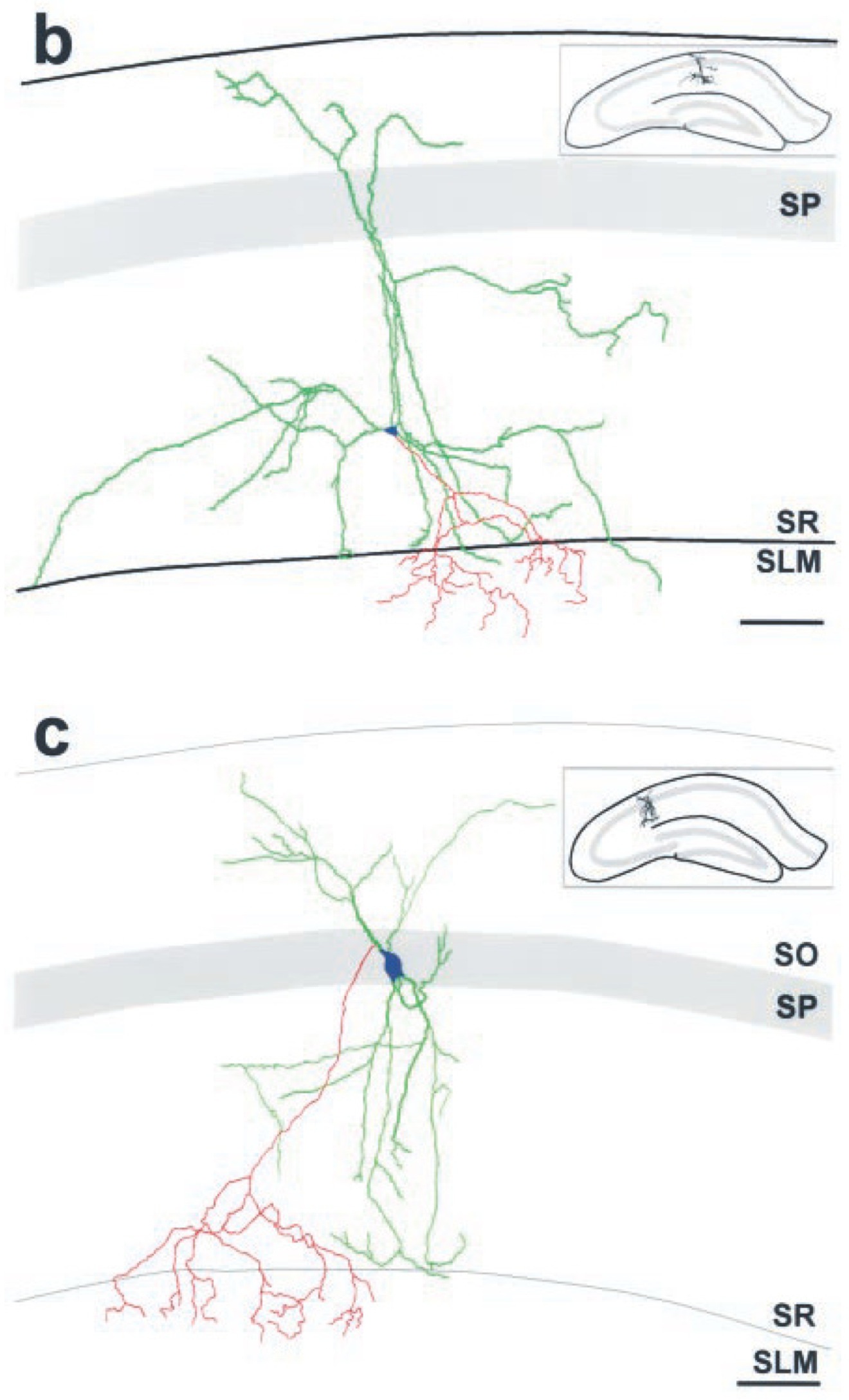
This name is a variant of "O-LM" since the dendrites are in SO and SR and the axons project to SLM. CA1 clarifies the subregion of origin, since the name is otherwise generic.

Synonym(s)
CA1 P-LM cell
CA1 R-LM cell
CA1 SR interneuron

List of articles
Click here to view the list
Oliva AA Jr  (2000) J Neurosci
Novel hippocampal interneuronal subtypes identified using transgenic mice that express green fluorescent protein in GABAergic interneurons.
Oliva AA Jr, Jiang M, Lam T, Smith KL, Swann JW.
J Neurosci, 2000 May 1, 20 (9), Pages: 3354 - 3368
PMID: 10777798
Tags: morphology, marker, membrane biophysics, firing patterns, synapse probabilities

Supplemental PMIDs
Click here to view the list of PMIDs

List of Hippocampome.org to NeuroMorpho.Org mappings
Click here to view the list of mappings
  • Esclapez

  • Morphology
    Soma
    CA1:SR
    CA1:SP
    Axons
    CA1:SLM
    Dendrites
    CA1:SR
    CA1:SO

    Representative figure
    Novel hippocampal interneuronal subtypes identified using transgenic mice that express green fluorescent protein in GABAergic interneurons.
    Oliva AA Jr, Jiang M, Lam T, Smith KL, Swann JW
    J Neurosci, 2000 May 1, 20 (9), pages: 3354 - 3368
    PMID: 10777798;


    "Figure 4. Neurolucida reconstructions of [CA1] EGFP[enhanced green fluorescent protein]-expressing O-LM [oriens lacunosum moleculare cells], R-LM [stratum radiatum interneurons with lacunosum-moleculare axon arborization], and P-LM [stratum pyramidale interneurons with lacunosum-moleculare axon arborization] cells, illustrating their complete in vivo dendritic structures and axonal innervation patterns. Reconstructions were performed on serial brain sections that were immunohistochemically processed for EGFP using DAB[diaminobenzidine]-based immunohistochemistry. Dendrites are green, axons are red, and somata are blue. É b, Reconstruction of an R-LM cell {CA1 OR-LM}. This cell had its somata in SR [stratum radiatum] and gave rise to an axon that ramified significantly in SLM [stratum lacunosum-moleculare]. This interneuronal subtype had dendrites that spanned from SO to SR, but rarely entered SLM. Note that one of the dendritic processes appears to deeply penetrate SLM; this is a misrepresentation attributable to the flattening of the three-dimensional reconstruction into two dimensions, in which laminar borders cannot properly be preserved. The dendritic tree of this R-LM cell extended ~400 [micro]m in the septotemporal axis and ~850 [micro]m in the mediolateral direction. c, Reconstruction of a P-LM cell {CA1 OR-LM}. This cell had its somata in SP and gave rise to an axon that ramified in SLM. As was the case for R-LM cells, this interneuronal subtype had dendrites that spanned from SO to SR and that tended to avoid all but the most proximal portion of SLM. The dendritic tree of this P-LM cell extended ~350 [micro]m in the septotemporal axis and ~400 [micro]m in the mediolateral direction. All reconstructions were from adult GIN mice. Note that only partial axonal reconstructions were possible: axons could only be followed for short distances after entering SLM because they became obscured by the high density of other EGFP-expressing axonal processes. Insets show the hippocampal location of each reconstructed interneuron. Strata abbreviations are the same as in Figure 2. Scale bars: a-c, 100 [micro]m."


    Molecular markers
    Positive
    AR-beta1 (multiple confirming inferences)
    mGluR1a (inference)
    SOM (confirmed by inference)
    Negative
    5HT-3 (inference)
    AR-beta2 (multiple confirming inferences)
    CR (inference)
    ENK (inference)
    ErbB4 (multiple confirming inferences)
    GABAa\alpha 6 (inference)
    MOR (multiple confirming inferences)
    PV (inference)
    vGluT3 (inference)
    Mixed expression
    None known

    Electrophysiological properties
    Key: [range] OR representative value±SD (measurements); Number of sources (total measurements): [min , max]

    Resting membrane potential (Vrest): [-69.0, -67.0] mV (2); 1 source
    Input resistance (Rin): [150.0, 300.0] MΩ (2); 1 source
    Time constant (τm): 68 ms (1); 1 source
    Threshold potential (Vthresh): [14.0, 16.2] mV (2); 1 source
    Fast afterhyperpolaziring potential amplitude (Fast AHP): [7.2, 9.6] mV (2); 1 source
    Action potential amplitude (APampl): [82.8, 89.2] mV (2); 1 source
    Slow afterhyperpolarizing potential amplitude (Slow AHP): 4.6 mV (1); 1 source
    Sag ratio: 0.71 (1); 1 source

    Notes
    Rejected merger with CA1 (-)3220 peforant path-associated (PPA) neurons, which is now merged into CA1 (-)3222 peforant path-associated (PPA) neurons. Related classes: CA1 (-)1002 oriens-lacunosum moleculare (O-LM) neurons (active) CA1 (-)1003 oriens-lacunosum moleculare (O-LM) neurons (active) CA1 (-)1102p-CA3 10000 oriens-lacunosum moleculare (O-LM) neurons (suspended) Cited by Somogyi & Klausberger 2005 but not picked up in their later papers. In Figure 4, borderline axons in SR are considered less than 15% since the authors report extensive branching in SLM but "axons could only be followed for short distances after entering SLM because they became obscured by the high density of other EGFP-expressing axonal processes."

    Firing Pattern
    Images
    Image MissingImage Missing
    Parameters View page
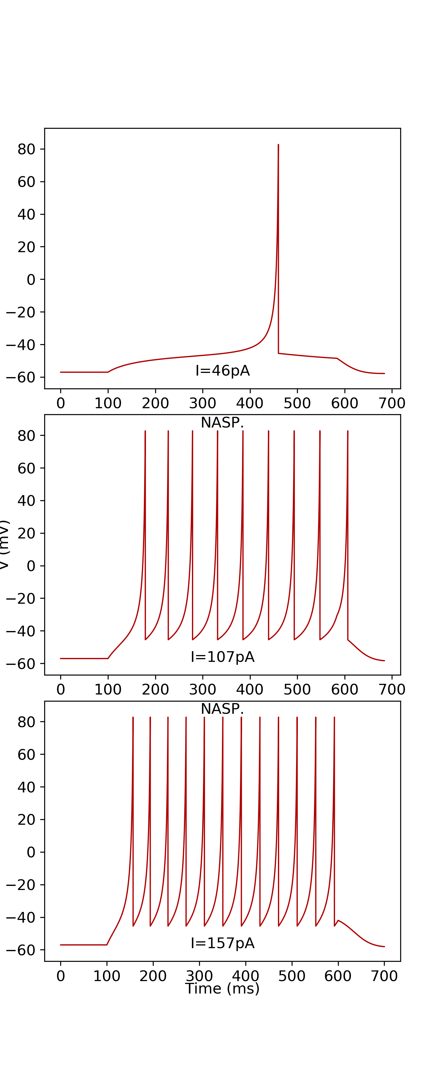
    NASP [Evidence] [Firing Pattern]

    Izhikevich Model
    Parameters Single Compartment Downloads Simulate
    subtype: 1 k=0.56;a= 0.014;b= 2.09;d= -15;C= 248;Vr= -57.03;Vt= -42.75;Vpeak= 82.73;Vmin= -45.49; Simulate
    Images
    Image Missing
    ModelDB Model
    Model Accession Number PubMed ID

    Sources of Input
    Known sources
    Excitatory or Inhibitory
    none known

    Potential sources
    Excitatory or Inhibitory
    CA3 Pyramidal
    CA3c Pyramidal
    CA3c Umbrella Pyramidal*
    CA3 Trilaminar
    CA2 Pyramidal
    CA2 Bistratified
    CA1 Pyramidal
    CA1 Deep Pyramidal
    CA1 Superficial Pyramidal
    CA1 Radiatum Giant
    CA1 Back-Projection
    CA1 Bistratified
    CA1 Hippocampo-subicular Projecting ENK+
    CA1 Ivy
    CA1 Interneuron Specific LM-R
    CA1 Interneuron Specific LMO-O
    CA1 LMR
    CA1 Interneuron Specific LMR-O*
    CA1 LMR-R*
    CA1 Interneuron Specific LMR-R
    CA1 LMR Projecting
    CA1 Recurrent O-LM
    CA1 O-LMR
    CA1 Recurrent O-LMR*
    CA1 Interneuron Specific O-R
    CA1 O-RP*
    CA1 Interneuron Specific O-Targeting QuadD
    CA1 Oriens/Alveus
    CA1 Oriens-Bistratified
    CA1 Oriens-QuadA Projecting
    CA1 O-RPO*
    CA1 QuadD-RP*
    CA1 Quadrilaminar
    CA1 Interneuron Specific R-O
    CA1 R-Receiving Apical-Targeting
    CA1 R-RO*
    CA1 R-RP*
    CA1 Radiatum
    CA1 Interneuron Specific RO-O
    CA1 Interneuron Specific RO-R*
    CA1 RO-RP*
    CA1 RP-RPO*
    CA1 Schaffer Collateral-Associated
    CA1 Schaffer Collateral-Correlated*
    CA1 Schaffer Collateral-Receiving R-Targeting
    CA1 SO-SO
    CA1 SO-SO Projecting*
    CA1 Trilaminar
    CA1 Radial Trilaminar
    Sub CA1-Projecting Pyramidal
    EC LIII Small Pyramidal
    EC LIII Pyramidal

    Potential sources known to be avoided
    Excitatory or Inhibitory
    CA1 Axo-axonic
    CA1 Horizontal Axo-axonic

    Targets of Output
    Known targets
    Excitatory or Inhibitory
    none known

    Potential targets
    Excitatory or Inhibitory
    CA1 Pyramidal
    CA1 Deep Pyramidal
    CA1 Superficial Pyramidal
    CA1 Cajal-Retzius
    CA1 Local-Projecting Cajal-Retzius*
    CA1 Radiatum Giant
    CA1 Axo-axonic
    CA1 Basket
    CA1 Vertical Basket*
    CA1 Basket CCK+
    CA1 Interneuron Specific LM-R
    CA1 Interneuron Specific LMO-O
    CA1 LMR
    CA1 LMR-LM*
    CA1 Interneuron Specific LMR-O*
    CA1 LMR-R*
    CA1 Interneuron Specific LMR-R
    CA1 LMR Projecting
    CA1 Neurogliaform
    CA1 Neurogliaform Projecting
    CA1 Interneuron Specific O-Targeting QuadD
    CA1 Oriens/Alveus
    CA1 Perforant Path-Associated
    CA1 Perforant Path-Associated QuadD
    CA1 QuadD-RP*
    CA1 Quadrilaminar
    CA1 Schaffer Collateral-Associated
    CA1 Radial Trilaminar

    Potential targets known to be avoided
    Excitatory or Inhibitory
    none known

    Oscillation Phase Locking
    Category
    Value