CA1 Horizontal Axo-axonic
* indicates new v2.0 neuron type
Name Supertype (ID:Family:Type:Subregion)
CA1 (i)0012 Horizontal Axo-axonic S110904:Perisomatic-targeting:Axo-axonic:CA1

Name derivation
This is the only name in the cited literature. CA1 distinguishes these cells from horizontal axo-axonic cells in other subregions.

Synonym(s)
CA1 horizontal axo-axonic cell

List of articles
Click here to view the list
Ganter P  (2004) Hippocampus
Properties of horizontal axo-axonic cells in stratum oriens of the hippocampal CA1 area of rats in vitro.
Ganter P, Szucs P, Paulsen O, Somogyi P.
Hippocampus, 2004, 14 (2), Pages: 232 - 243
PMID: 15098728; DOI: 10.1002/hipo.10170
Tags: morphology, membrane biophysics, connectivity, synapse probabilities
Martinez A  (1996) J Comp Neurol
Regional variability and postsynaptic targets of chandelier cells in the hippocampal formation of the rat.
Martinez A, Lubke J, Del Rio JA, Soriano E, Frotscher M.
J Comp Neurol, 1996 Dec 02, 376 (1), Pages: 28 - 44
PMID: 8946282; DOI: 10.1002/(SICI)1096-9861(19961202)376:1<28::AID-CNE2>3.0.CO;2-Q
Tags: synapse probabilities

Supplemental PMIDs
Click here to view the list of PMIDs


Morphology
Soma
CA1:SO
Axons
CA1:SP
Dendrites
CA1:SO

Representative figure
Properties of horizontal axo-axonic cells in stratum oriens of the hippocampal CA1 area of rats in vitro.
Ganter P, Szucs P, Paulsen O, Somogyi P
Hippocampus, 2004, 14 (2), pages: 232 - 243
PMID: 15098728; DOI: 10.1002/hipo.10170


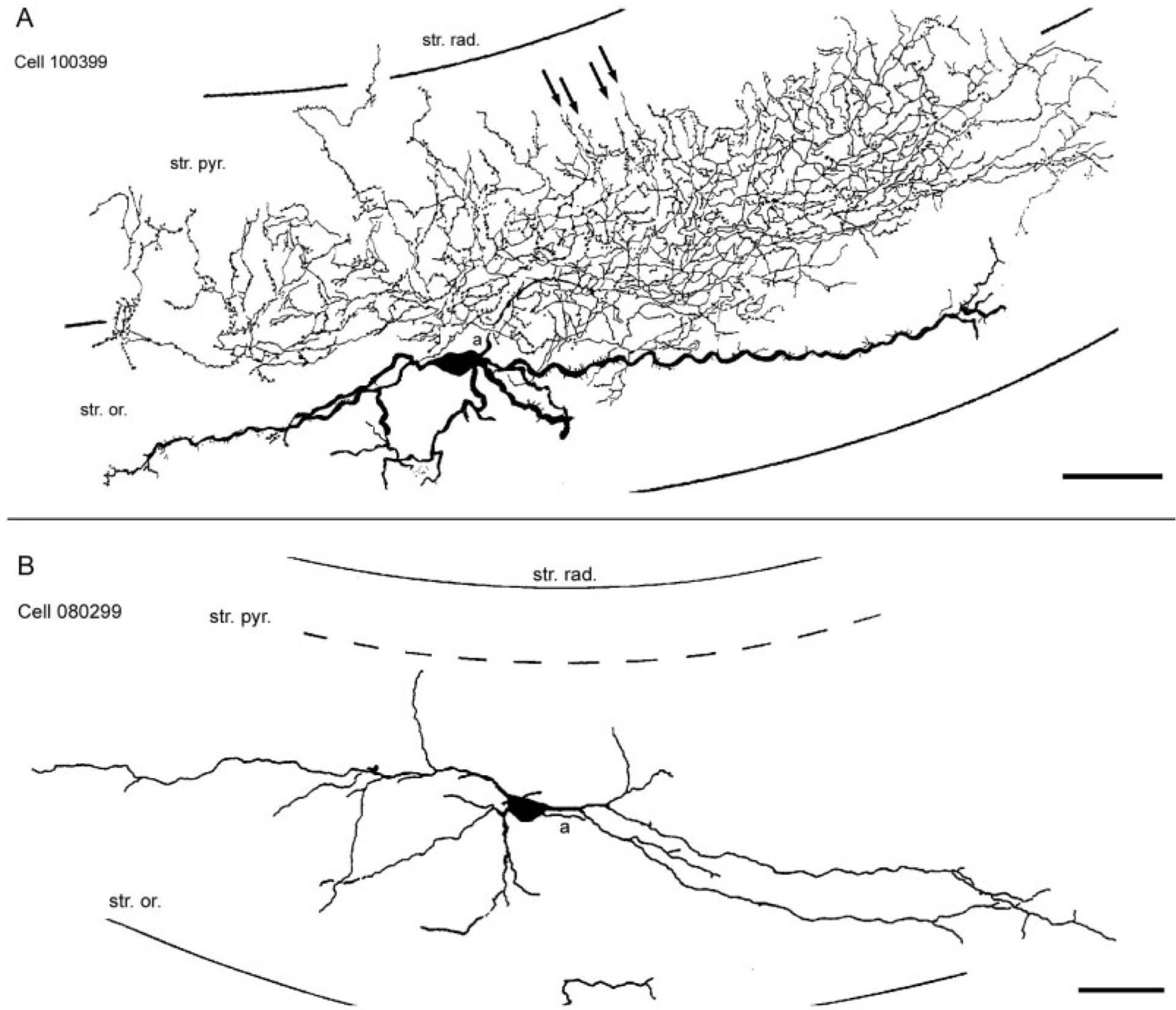
"FIGURE 1. Identification of [CA1] horizontal axo-axonic cells {CA1 Horizontal Axo-axonic}. A: Reconstruction of a cell showing dendrites (thick processes) running mainly parallel with the layers in stratum oriens (str. or.) and axonal arbor mostly in the deep pyramidal layer (str. pyr.) and adjacent stratum oriens. Note the characteristic radial axonal terminal segments (arrows) defining it as an axo-axonic cell. Some shrinkage is likely to have occurred during the histological processing, which is apparent from the undulating appearance of the dendrites. B: Reconstruction of the dendritic tree of another cell also shown in C-F. The broken line denotes the compact sublayer of stratum pyramidale, as also seen in the micrograph shown in E. C: Light micrograph of the same cell shown together with a reciprocally connected pyramidal cell (P). The reciprocal connection was evident from the synaptic responses (Figs. 4A and 5)."


Molecular markers
Positive
Gaba-a-alpha (multiple confirming inferences)
PV
Negative
5HT-3 (confirmed by inference)
AR-beta2 (inference)
CB (multiple confirming inferences)
CB1 (multiple confirming inferences)
CCK
Chrna2 (inference)
CoupTF II
CR (confirmed by inference)
ENK (confirmed by inference)
GABAa\alpha 6 (inference)
mGluR1 (inference)
mGluR1a (inference)
MOR (inference)
nNOS
NPY (confirmed by inference)
SOM (confirmed by inference)
vGluT3 (confirmed by inference)
VIP (confirmed by inference)
Mixed expression
None known

Electrophysiological properties
Key: [range] OR representative value±SD (measurements); Number of sources (total measurements): [min , max]

Input resistance (Rin): [186.0, 252.0] MΩ (2); 1 source
Time constant (τm): [16.0, 32.0] ms (2); 1 source
Fast afterhyperpolaziring potential amplitude (Fast AHP): [6.0, 13.7] mV (2); 1 source
Action potential amplitude (APampl): [71.4, 90.2] mV (2); 1 source
Action potential width (APwidth): [0.7, 1.1] ms (2); 1 source
Max firing rate (Max F.R.): 87 Hz (1); 1 source
Resting membrane potential (Vrest): -57±5 mV (15); 1 source
Threshold potential (Vthresh): 25 mV (1); 1 source
Slow afterhyperpolarizing potential amplitude (Slow AHP): 6.1 mV (1); 1 source
Sag ratio: 0.74 (1); 1 source

Notes
Axons exclusively target the axon initial segment of CA1 pyramidal cells (and, unlike basket cells, do not target other interneurons) perisomatic axonal targeting interpreted as PCL extended interpretation of SP in apical and basal directions

Firing Pattern
Images
Image Missing
Parameters View page
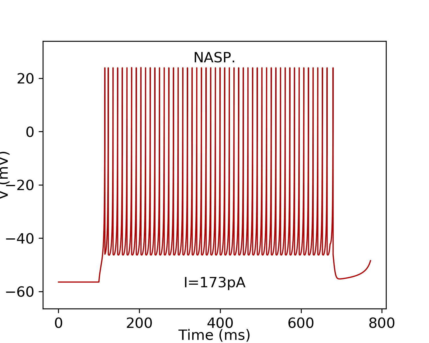
NASP [Evidence] [Firing Pattern]

Izhikevich Model
Parameters Single Compartment Downloads Simulate
subtype: 1 k=2.15;a= 0.03;b= -47.85;d= 277;C= 88;Vr= -56.45;Vt= -41.32;Vpeak= 23.89;Vmin= -46.01; Simulate
Images
Image Missing
ModelDB Model
Model Accession Number PubMed ID

Sources of Input
Known sources
Excitatory or Inhibitory
CA1 Pyramidal

Potential sources
Excitatory or Inhibitory
CA3 Pyramidal
CA3c Pyramidal
CA3 Trilaminar
CA2 Pyramidal
CA2 Bistratified
CA1 Deep Pyramidal
CA1 Superficial Pyramidal
CA1 Radiatum Giant
CA1 Back-Projection
CA1 Bistratified
CA1 Hippocampo-subicular Projecting ENK+
CA1 Ivy
CA1 Interneuron Specific LMO-O
CA1 Interneuron Specific LMR-O*
CA1 Recurrent O-LM
CA1 Recurrent O-LMR*
CA1 Interneuron Specific O-Targeting QuadD
CA1 Oriens/Alveus
CA1 Oriens-Bistratified
CA1 Oriens-QuadA Projecting
CA1 O-RPO*
CA1 Quadrilaminar
CA1 Interneuron Specific R-O
CA1 R-RO*
CA1 Interneuron Specific RO-O
CA1 RP-RPO*
CA1 Schaffer Collateral-Associated
CA1 Schaffer Collateral-Correlated*
CA1 SO-SO
CA1 SO-SO Projecting*
CA1 Trilaminar
CA1 Radial Trilaminar
EC LIII Small Pyramidal
EC LIII Pyramidal

Potential sources known to be avoided
Excitatory or Inhibitory
none known

Targets of Output
Known targets
Excitatory or Inhibitory
CA1 Pyramidal

Potential targets
Excitatory or Inhibitory
CA2 Wide-Arbor Basket
CA1 Deep Pyramidal
CA1 Superficial Pyramidal
CA1 Back-Projection
CA1 Vertical Basket*
CA1 Oriens/Alveus
CA1 Perforant Path-Associated QuadD
CA1 Quadrilaminar
CA1 RP-RPO*

Potential targets known to be avoided
Excitatory or Inhibitory
CA1 Axo-axonic
CA1 Basket
CA1 Horizontal Basket
CA1 R-P Basket*
CA1 Trilayer Basket*
CA1 Basket CCK+
CA1 Trilayer Basket CCK+*
CA1 Bistratified
CA1 Ivy
CA1 Interneuron Specific LMO-O
CA1 Interneuron Specific LMR-R
CA1 Interneuron Specific O-Targeting QuadD
CA1 OR-LM
CA1 QuadD-RP*
CA1 Interneuron Specific R-O
CA1 Interneuron Specific RO-O
CA1 Radial Trilaminar

Oscillation Phase Locking
Category
Value