|
|
|
* indicates new v2.0 neuron type
|
|
Name
|
|
Supertype (ID:Family:Type:Subregion)
|
|
|
CA1 (i)3000 Neurogliaform
|
S101304:Ivy/Neurogliaform Family:Ivy/Neurogliaform:CA1
|
|
Name derivation
|
|
|
|
This is the only name in the cited literature and is the canonical CA1 neurogliaform cell. CA1 distinguishes these cells from neurogliaform cells in other subregions.
|
|
Synonym(s)
|
|
|
|
CA1 neurogliaform cell
|
|
|
CA1 neurogliaform neuron
|
|
|
CA1 NG
|
|
|
CA1 NG cell
|
|
|
CA1 NGFC
|
|
|
Click here to view the list
Bartos M (2010) Hippocampal Microcircuits: A Computational Modeler's Resource Book
Fast and Slow GABAergic Transmission in Hippocampal Circuits Bartos M, Sauer JF, Vida I, Kulik A.
Hippocampal Microcircuits: A Computational Modeler's Resource Book,
2010,
Pages: 129 - 162
ISBN: 9781441909954
Tags: morphology, connectivity, synapse probabilities
Fuentealba P (2008) Neuron
Ivy cells: a population of nitric-oxide-producing, slow-spiking GABAergic neurons and their involvement in hippocampal network activity. Fuentealba P, Begum R, Capogna M, Jinno S, Marton LF, Csicsvari J, Thomson A, Somogyi P, Klausberger T.
Neuron,
2008 Mar 27, 57 (6),
Pages: 917 - 929
PMID: 18367092; DOI: 10.1016/j.neuron.2008.01.034
Tags: morphology, connectivity, phases, synapse probabilities
Gloveli T (2010) Hippocampal Microcircuits: A Computational Modeler's Resource Book
Neuronal Activity Patterns During Hippocampal Network Oscillations In Vitro Gloveli T, Kopell N, Dugladze T.
Hippocampal Microcircuits: A Computational Modeler's Resource Book,
2010,
Pages: 247 - 276
ISBN: 9781441909954
Tags: morphology, connectivity, synapse probabilities
Karayannis T (2010) J Neurosci
Slow GABA transient and receptor desensitization shape synaptic responses evoked by hippocampal neurogliaform cells. Karayannis T, Elfant D, Huerta-Ocampo I, Teki S, Scott RS, Rusakov DA, Jones MV, Capogna M.
J Neurosci,
2010 Jul 21, 30 (29),
Pages: 9898 - 9909
PMID: 20660272; DOI: 10.1523/JNEUROSCI.5883-09.2010
Tags: morphology, connectivity, synapse probabilities
Klausberger T (2008) Science
Neuronal diversity and temporal dynamics: the unity of hippocampal circuit operations. Klausberger T, Somogyi P.
Science,
2008 Jul 4, 321 (5885),
Pages: 53 - 57
PMID: 18599766; DOI: 10.1126/science.1149381
Tags: morphology, connectivity, synapse probabilities
Klausberger T (2009) Eur J Neurosci
GABAergic interneurons targeting dendrites of pyramidal cells in the CA1 area of the hippocampus. Klausberger T.
Eur J Neurosci,
2009 Sep, 30 (6),
Pages: 947 - 957
PMID: 19735288; DOI: 10.1111/j.1460-9568.2009.06913.x
Tags: morphology, marker, connectivity, synapse probabilities
Price CJ (2005) J Neurosci
Neurogliaform neurons form a novel inhibitory network in the hippocampal CA1 area. Price CJ, Cauli B, Kovacs ER, Kulik A, Lambolez B, Shigemoto R, Capogna M.
J Neurosci,
2005 Jul 20, 25 (29),
Pages: 6775 - 6786
PMID: 16033887; DOI: 10.1523/JNEUROSCI.1135-05.2005
Tags: morphology, marker, membrane biophysics, connectivity, firing patterns, synapse probabilities
Price CJ (2008) J Neurosci
GABA(B) receptor modulation of feedforward inhibition through hippocampal neurogliaform cells. Price CJ, Scott R, Rusakov DA, Capogna M.
J Neurosci,
2008 Jul 2, 28 (27),
Pages: 6974 - 6982
PMID: 18596171; DOI: 10.1523/JNEUROSCI.4673-07.2008
Tags: morphology, connectivity, synapse probabilities
Sakalar E (2022) Science
Neurogliaform cells dynamically decouple neuronal synchrony between brain areas Sakalar E, Klausberger T, Lasztoczi B.
Science,
2022, 377 (6603),
Pages: 324 - 328
PMID: 35857593; DOI: 10.1126/science.abo3355
Tags: phases
Somogyi P (2010) Handbook of Brain Microcircuits
Chapter: Hippocampus: Intrinsic organisation Somogyi P.
Handbook of Brain Microcircuits,
2010,
Pages: 148 - 164
ISBN: 9780195389883
Tags: morphology, connectivity, synapse probabilities
|
|
Supplemental PMIDs
|
|
|
|
Click here to view the list of PMIDs
|
|
List of Hippocampome.org to NeuroMorpho.Org mappings
|
|
|
Click here to view the list of mappings
|
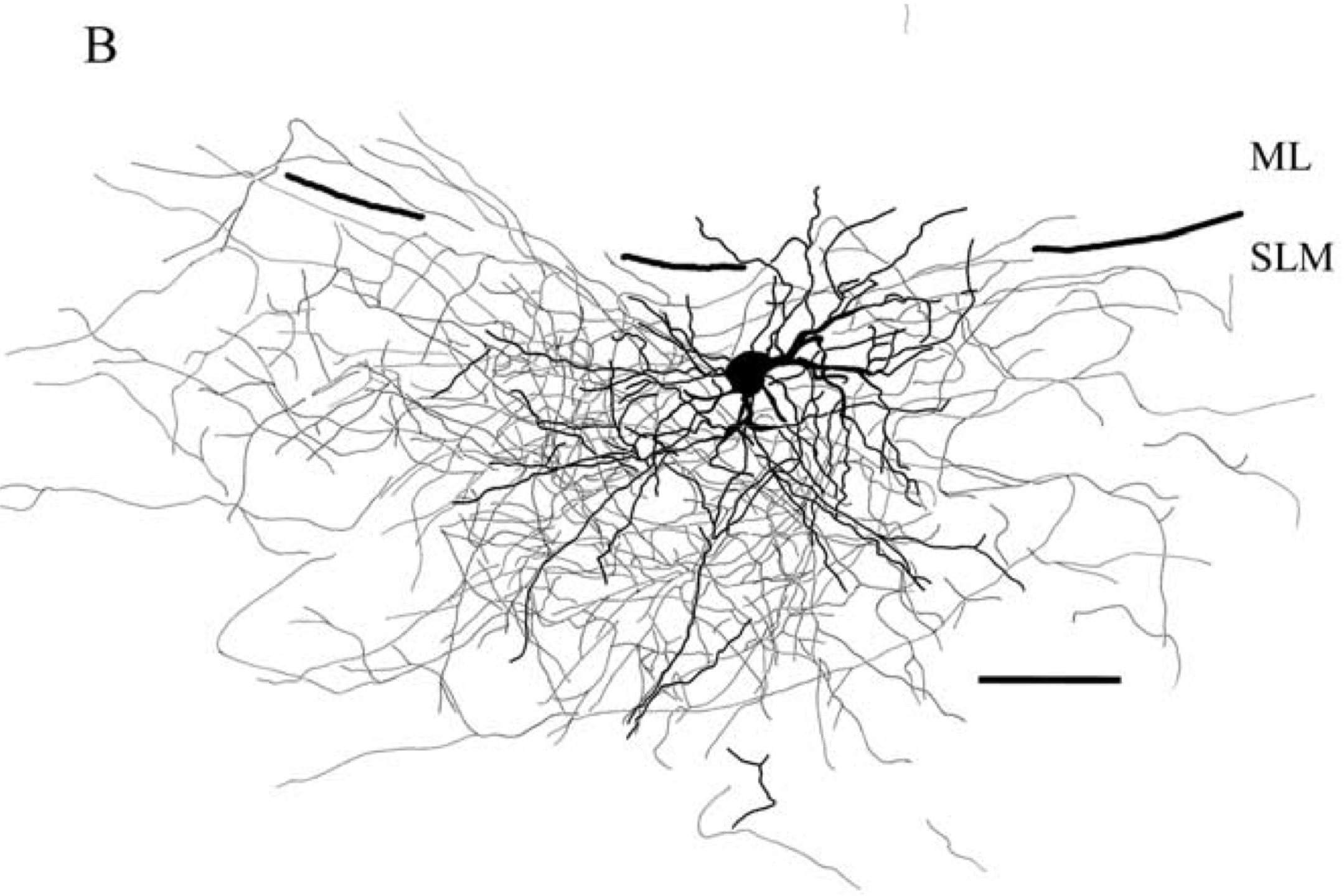
"Figure 1. Reconstruction of NG [neurogliaform] cells in the SLM [stratum lacunosum-moleculare] of area CA1. ... B, Reconstruction of another NG cell {CA1 Neurogliaform} (P13) illustrating dendrites (black) and axons (gray). Perforant pathway stimulation at 40 Hz evoked EPSCs with a depressing pattern in this cell, like the pattern shown in Figure 4 A1. The dendrites and axon are mostly restricted to the CA1 SLM. SLM, Stratum lacunosum moleculare; ML, molecular layer of the dentate gyrus." |
|
Electrophysiological properties
|
| Key: [range] OR representative value±SD (measurements); Number of sources (total measurements): [min , max] | |
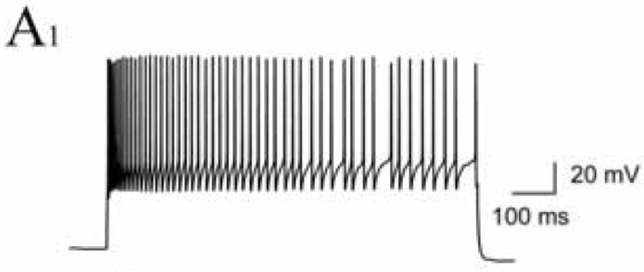
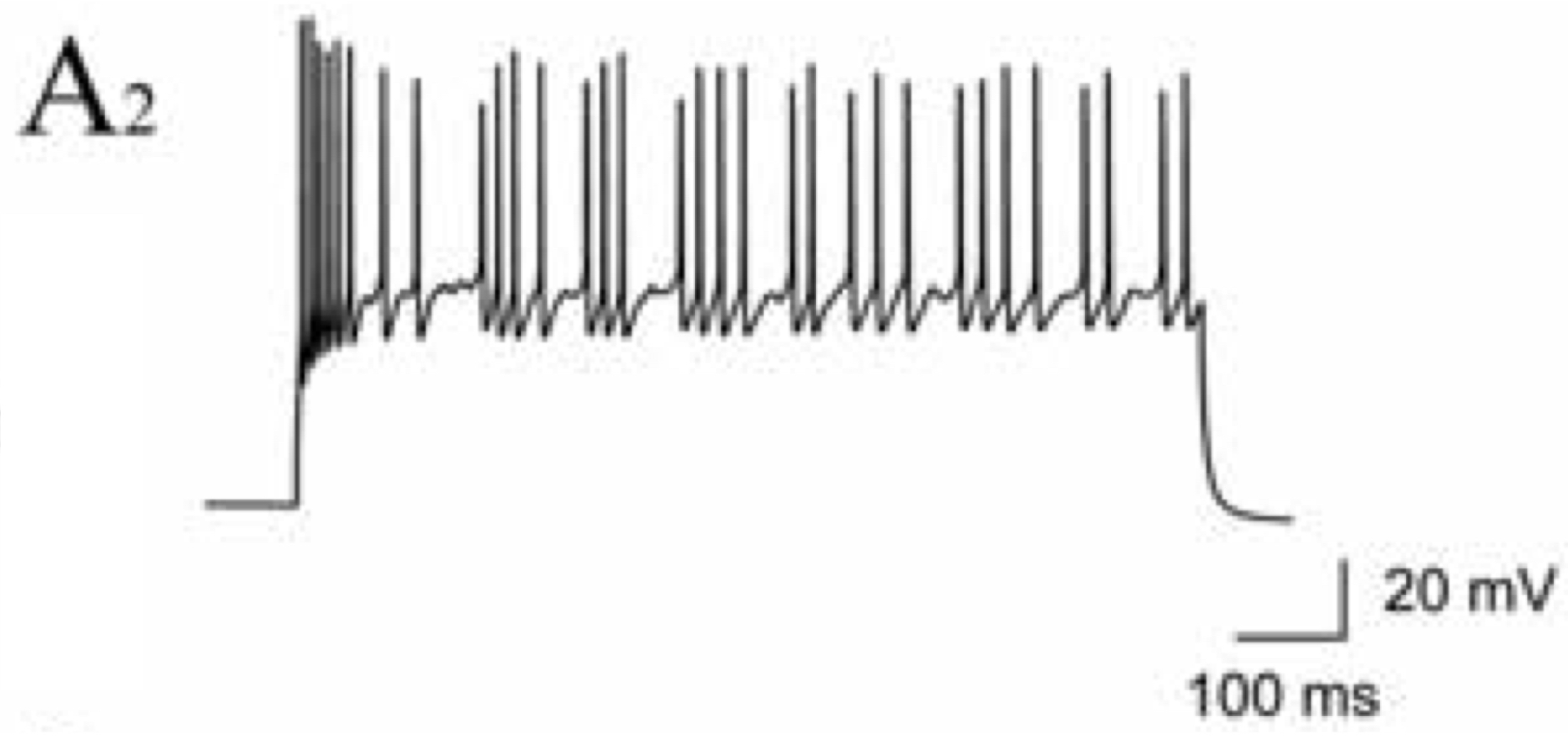
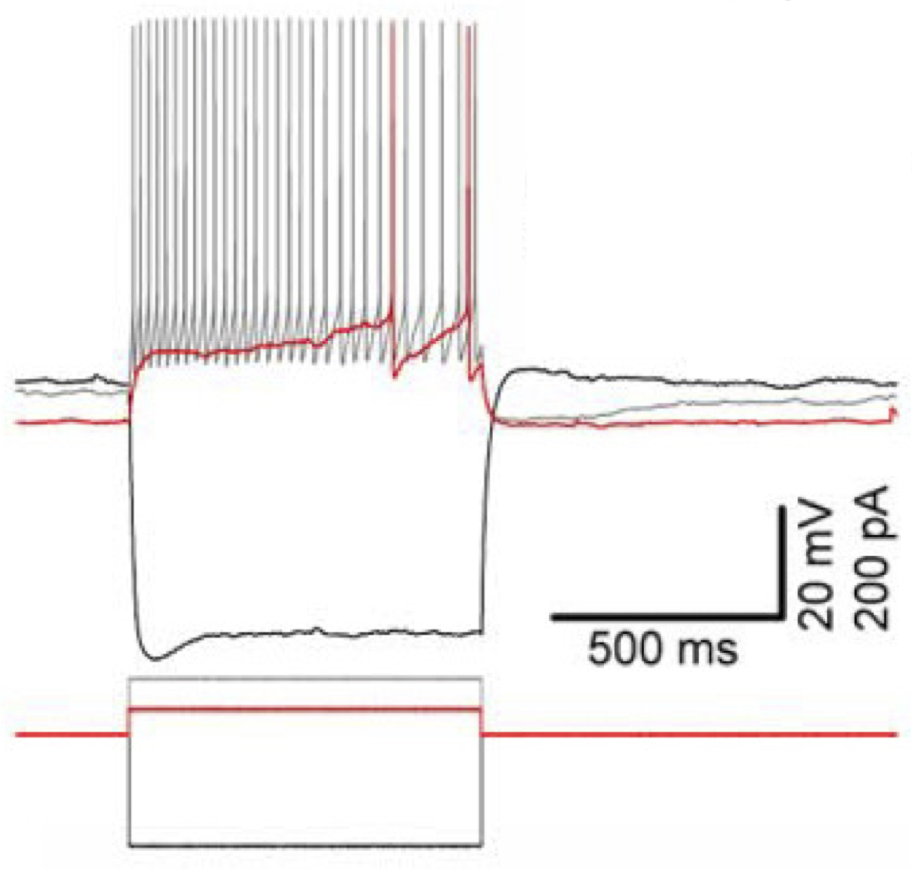
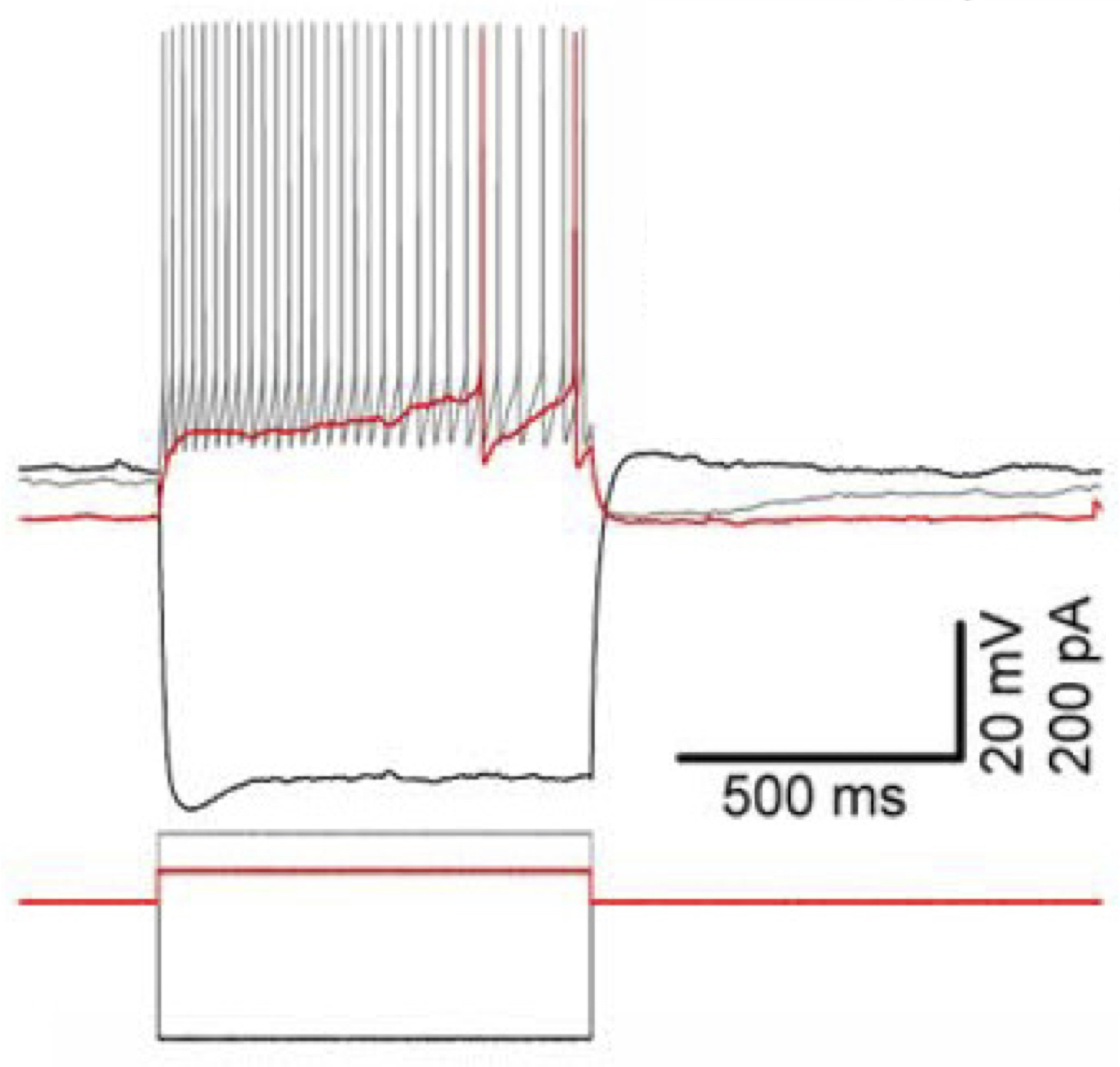
| | Resting membrane potential (Vrest): -63.1±5.6 mV (33); 2 sources (67): [-70 , -63.1] | | Input resistance (Rin): 215.3±92.8 MΩ (32); 2 sources (66): [215.3 , 302] | | Time constant (τm): 12.43±4.59 ms (32); 2 sources (66): [12.43 , 25] | | Threshold potential (Vthresh): 32.4 mV (1); 2 sources (2): [32.4 , 36] | | Fast afterhyperpolaziring potential amplitude (Fast AHP): 20.4±4.1 mV (34); 2 sources (68): [20.4 , 21] | | Action potential amplitude (APampl): 73 mV (1); 2 sources (35): [52 , 73] | | Action potential width (APwidth): 0.9±0.18 ms (26); 2 sources (60): [0.9 , 1.08] | | Max firing rate (Max F.R.): >60 Hz (1); 2 sources (27): [52.8 , 60] | | Slow afterhyperpolarizing potential amplitude (Slow AHP): 9 mV (1); 2 sources (2): [5.5 , 9] | | Sag ratio: 0.99 (1); 2 sources (35): [0.88 , 0.99] |
| Parameters
|
Single Compartment
|
Downloads
|
Simulate
|
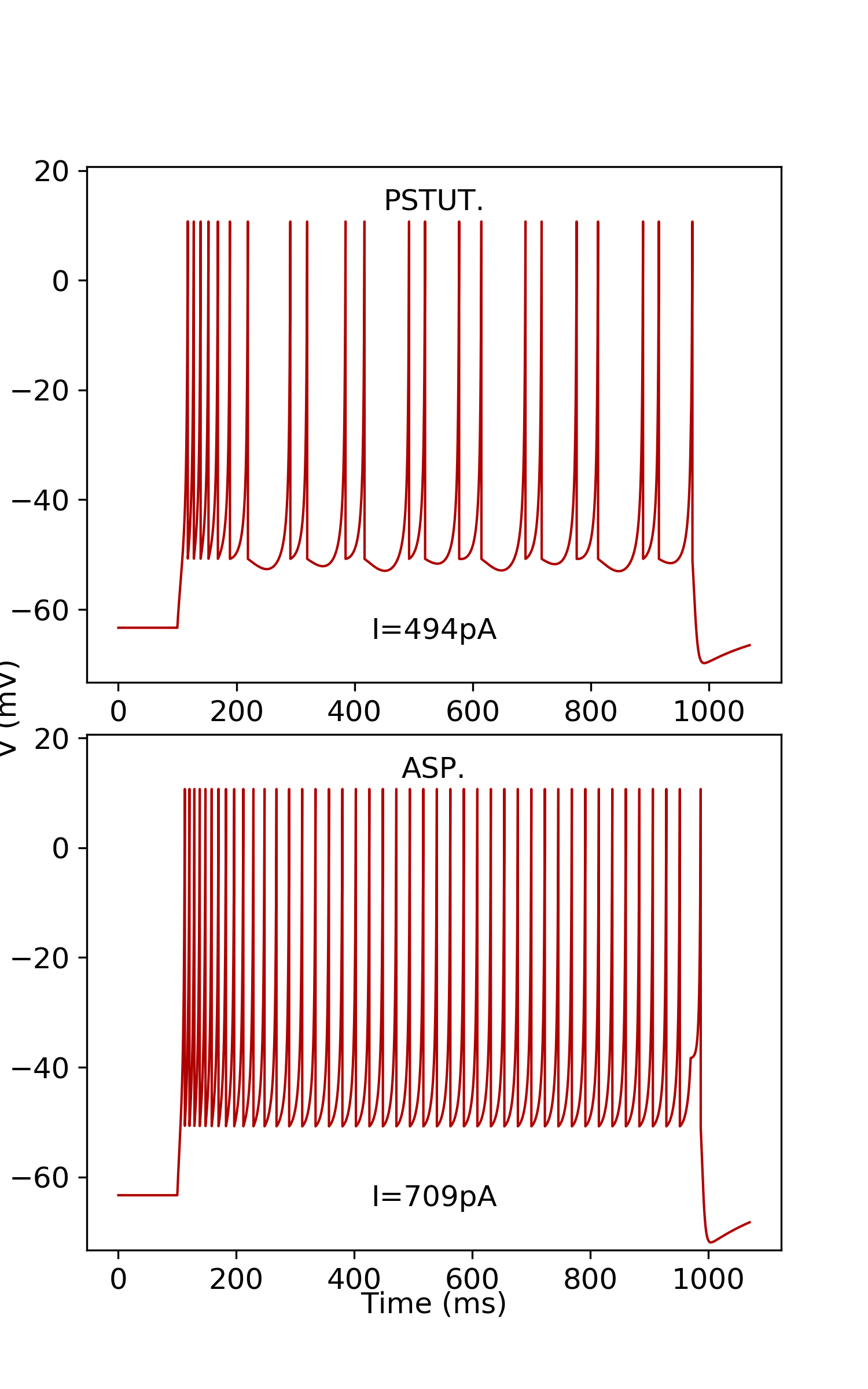
| subtype: 1 k=2.36;a= 0.009;b= 17.56;d= 40;C= 254;Vr= -63.33;Vt= -47.62;Vpeak= 10.7;Vmin= -50.78; | | Simulate
|
|
|
|
|
Potential sources known to be avoided
|
|
|
|
|
|
Potential targets known to be avoided
|
|
| Oscillation Phase Locking |
| Category | Value | | Theta (deg): | 194.00 | | Theta range: | [177.00, 215.00] | | Theta measurements: | 5 | | Theta representative selection: | mice, adult, male,
juxtacellular, head-fixed awake | | In vivo firing rate (Hz): | 3.40 | | Firing rate range: | [3.40, 3.40] | | Firing rate measurements: | 1 | | Firing rate representative selection: | mice, adult, male,
juxtacellular, head-fixed awake | | Other: | gamma and 1 other | | Other representative selection: | mice, adult, male,
juxtacellular, head-fixed awake |
|
 Browse
 Search
 Help