|
| Morphology |
| Soma | |
| CA1:SLM |
| Axons | |
| DG:SMo | |
| CA1:SLM |
| Dendrites | |
| DG:SMo | |
| CA1:SLM |
| Representative figure |
|
Neurogliaform neurons form a novel inhibitory network in the hippocampal CA1 area. Price CJ, Cauli B, Kovacs ER, Kulik A, Lambolez B, Shigemoto R, Capogna M J Neurosci, 2005 Jul 20, 25 (29), pages: 6775 - 6786 PMID: 16033887; DOI: 10.1523/JNEUROSCI.1135-05.2005 |
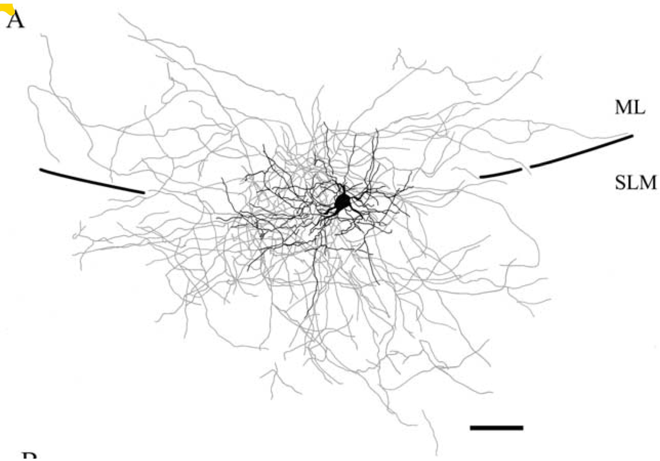 "Figure 1. Reconstruction of NG [neurogliaform] cells in the SLM [stratum lacunosum-moleculare] of area CA1. A, Reconstruction of an NG cell {CA1 Neurogliaform Projecting} (P18) illustrating dendrites (black) and axons (gray). Perforant pathway stimulation at 40 Hz elicited EPSCs [excitatory postsynaptic currents] with a facilitating/depressing pattern in this cell, like the pattern shown in Figure 4 A2. Note that the dendrites and axons cross the hippocampal fissure and extend both into the molecular layer of the dentate gyrus and the SLM of area CA1." |
| Molecular markers |
| Positive | |
| alpha-actinin-2 | |
| CoupTF II (confirmed by inference) | |
| GABA-B1 | |
| Gaba-a-alpha (confirmed by inference) | |
| NPY | |
| RLN (confirmed by inference) |
| Negative | |
| AR-beta1 (inference) | |
| AR-beta2 (inference) | |
| CB (confirmed by inference) | |
| DYN | |
| GABAa\alpha 6 (inference) | |
| MOR | |
| PV (confirmed by inference) | |
| vGluT3 | |
| VIP (confirmed by inference) |
| Mixed expression | |
| CCK (subtypes) | |
| CR (subtypes) | |
| ENK (subtypes) | |
| nNOS (subtypes) | |
| SOM (subtypes) | |
| Electrophysiological properties |
| Key: [range] OR representative value±SD (measurements); Number of sources (total measurements): [min , max] | |
| Resting membrane potential (Vrest): -63.1±5.6 mV (33); 1 source | |
| Input resistance (Rin): 215.3±92.8 MΩ (32); 1 source | |
| Time constant (τm): 12.43±4.59 ms (32); 1 source | |
| Threshold potential (Vthresh): [37.5, 51.3] mV (2); 2 sources (3): [32.4 , 37.5] | |
| Fast afterhyperpolaziring potential amplitude (Fast AHP): [14.9, 20.3]±4.1 mV (34); 2 sources (36): [14.9 , 20.4] | |
| Action potential amplitude (APampl): [32.0, 61.0] mV (2); 2 sources (3): [32 , 73] | |
| Action potential width (APwidth): 0.9±0.18 ms (26); 1 source | |
| Max firing rate (Max F.R.): 52.8±31 Hz (26); 1 source | |
| Slow afterhyperpolarizing potential amplitude (Slow AHP): 9 mV (1); 2 sources (2): [2.4 , 9] | |
| Sag ratio: 0.99 (1); 2 sources (2): [0.97 , 0.99] |
| Firing Pattern |
| Images | |
   |
| Parameters | View page | |
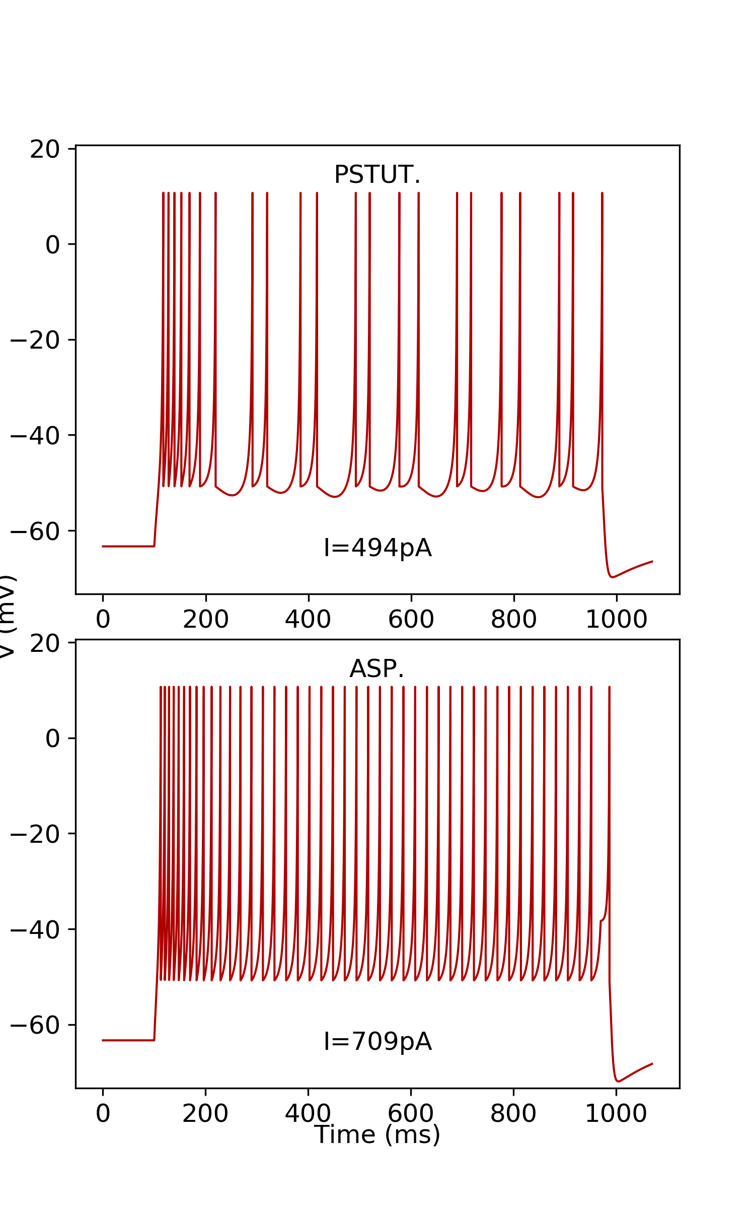
| ASP. | [Evidence] [Firing Pattern] | |
| PSTUT | [Evidence] [Firing Pattern] |
| Izhikevich Model |
| Parameters | Single Compartment | Downloads | Simulate |
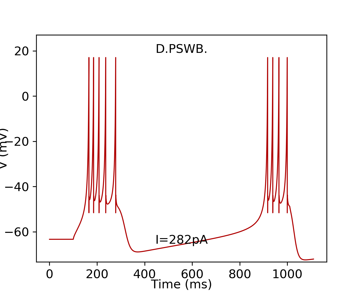
| subtype: 1 k=1.86;a= 0.003;b= -2.72;d= 90;C= 456;Vr= -63.35;Vt= -56.26;Vpeak= 17.06;Vmin= -52.77; | Simulate | ||
| subtype: 2 k=2.36;a= 0.009;b= 17.56;d= 40;C= 254;Vr= -63.33;Vt= -47.62;Vpeak= 10.7;Vmin= -50.78; | Simulate |
| Images | |
  |
| ModelDB Model |
| Model Accession Number | PubMed ID |
| Sources of Input |
| Targets of Output |
|
|
|
| Oscillation Phase Locking |
| Theta (deg): | 12.00 |
| Theta range: | [12.00, 19.00] |
| Theta measurements: | 2 |
| Theta representative selection: | rats, adult, unknown sex, juxtacellular, urethane plus supplemental doses of ketamine and xylazine |
| SWR ratio: | 0.27 |
| SWR ratio range: | [0.27, 1.11] |
| SWR ratio measurements: | 2 |
| SWR ratio representative selection: | rats, adult, unknown sex, juxtacellular, urethane plus supplemental doses of ketamine and xylazine |
| In vivo firing rate (Hz): | 1.50 |
| Firing rate range: | [1.50, 3.80] |
| Firing rate measurements: | 2 |
| Firing rate representative selection: | rats, adult, unknown sex, juxtacellular, urethane plus supplemental doses of ketamine and xylazine |
| Other: | firingRateNonBaseline |
| Other representative selection: | rats, adult, unknown sex, juxtacellular, urethane plus supplemental doses of ketamine and xylazine |
 Browse
 Search
 Help